A 33-year-old woman with an anomalous left coronary artery arising from the pulmonary artery who had undergone Takeuchi repair at age 7 years presented for evaluation. The Takeuchi procedure creates an aortopulmonary window and an intrapulmonary tunnel that baffles the left coronary artery to the aorta. A mediastinal mass was identified as a giant aneurysm of the left coronary artery resulting in compression of the pulmonary artery and left upper pulmonary vein. The patient underwent open repair with patch closure at the aortic entrance of the left coronary Takeuchi repair and resection and evacuation of the aneurysm. A saphenous vein graft to the left anterior descending artery was performed. Postoperative echocardiography demonstrated normal left ventricular function. This is the first reported case of giant aneurysm formation after Takeuchi repair. The reported complications have included the development of pulmonary artery stenosis at the intrapulmonary baffle, baffle leak, decreased left ventricular function, and mitral regurgitation. In conclusion, late complications of the Takeuchi procedure are common, underscoring the importance of lifelong follow-up at a center with experience in treating coronary anomalies.
Anomalous left coronary artery from the pulmonary artery (ALCAPA) is a rare, but serious, congenital coronary abnormality first reported in 1885 by Brooks. It generally presents in infancy with features of cardiac ischemia or heart failure, although it can present as late as adulthood. Because ALCAPA is associated with both morbidity and mortality, early surgical repair is recommended. However, given the evolution in surgical strategies during the past several decades, little is known about the types of complications that can occur later in life after ALCAPA repair.
Case Report
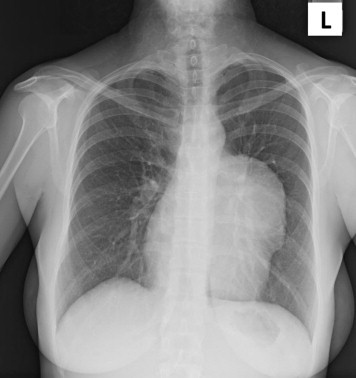
A 33-year-old woman was found to have ALCAPA at age 7 years and underwent repair with the Takeuchi procedure, without any reported complications. After the repair, she was followed up regularly as a child and was unaware of any abnormalities. As an adult, she had not received regular care from a cardiologist. She had successfully carried a twin pregnancy to full gestation without complications at age 31. In the year before presentation, she began experiencing palpitations and dyspnea on exertion and sought a cardiovascular evaluation. The physical examination disclosed a soft precordial systolic ejection murmur. The chest radiograph showed a mediastinal mass abutting the left cardiac contour ( Figure 1 ). Occasional ventricular premature complexes were seen on the electrocardiogram and by Holter monitoring. An echocardiogram revealed a large vascular mass overlying the main pulmonary artery that was partially thrombosed. The left ventricular size and ejection fraction were normal, with no regional wall motion abnormalities. Chest computed tomography identified the mass as a giant (9 × 8 cm) aneurysm of the left main coronary artery ( Figure 2 ). The aneurysm had a small patent lumen but was otherwise thrombosed ( Figure 3 ). It had caused external compression of the pulmonary trunk and left upper pulmonary vein. A chest radiograph at age 17 years had disclosed prominence of the left heart border.