(1)
Ospedale Civile di Vigevano, Vigevano, Italy
An Anatomical Overview of the Excitation-Conduction System of the Heart
The electrical activity of the heart is governed by the sinoatrial (SA) node (also known as the node of Keith and Flack) (Fig. 1.1), a microscopic structure located in the right atrium, at the junction between the atrium and the superior vena cava. It is made of pacemaker cells, which are intrinsically capable of generating rhythmic electrical impulses at rates that normally range from 60 to 100 beats per minute (bpm). The electrical current that originates here is propagated to the atria along preformed pathways known as the internodal tracts: the anterior internodal tract (known as Bachmann’s bundle), which includes a branch for the left atrium; the middle internodal tract (Wenckebach’s bundle); and the posterior internodal tract (Thorel’s bundle).
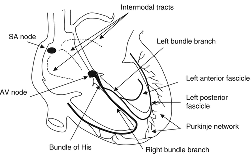
Fig. 1.1
The anatomy of the conduction system. SA sinoatrial, AV atrioventricular. See text for details
From the atria, the depolarization wave spreads to the atrioventricular (AV) node (or node of Tawara), the second fundamental station in the heart’s electrical conduction system. It is located in the posteroinferior region of the interatrial septum near the opening of the coronary sinus and represents the only normal connection between the atria and the ventricles. It is therefore the obligatory point of passage for impulses travelling to the ventricles.
Propagation of the electrical impulses from the AV node to the ventricles themselves occurs through a specialized conduction system. The proximal portion, which is known as the bundle of His, begins at the AV node and then divides within the ventricular septum to form the right and left bundle branches. The right bundle branch continues down the right side of the septum, just beneath the endocardium, and at the base of the anterior papillary muscle in the right ventricle, it divides again, sending fibers to the free wall of the right ventricle and to the left side of the septum. The left bundle branch, which is larger in caliber than the right, divides to form an anterior fascicle, which supplies the wall of the left ventricle, and an posterior fascicle, which supplies the left side of the septum. The two bundle branches continue to divide, forming a densely ramified subendocardial system known as the Purkinje fiber network, which propagates the depolarization current throughout the ventricular myocardium.
The Physiology of Impulse Formation and Conduction
The SA node has an intrinsic discharge rate (normal range: 60–100 bpm) that is higher than those of other parts of the myocardium. The AV node, which is the most important subsidiary station in the conduction system, emits impulses at a slightly lower rate (40–60 bpm). Under normal and pathological conditions, other portions of the conduction system or even the ordinary working myocardium are capable of firing at rates ranging from 20 to 40 bpm (Fig. 1.2).
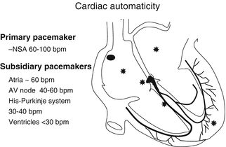
Fig. 1.2
The hierarchy of cardiac pacemakers. Asterisks indicate areas of the myocardium itself that are potential ectopic foci
Contraction of the heart is regulated by a continuous process of repetitive electrical excitation of the myocardium in which every electrical event is followed by a mechanical event.
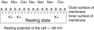
The electrical and contractile activities of the heart are mediated by the constant flow of ions (mainly sodium, calcium, and potassium) through lipoprotein structures in the cardiomyocyte cell membrane known as ion channels. If microelectrodes were placed on both sides of this membrane, it would show that, under resting conditions, the inside of the cell has a negative charge while the outside is positively charged. The result is a negative resting transmembrane potential of approximately −90 millivolts) (Fig. 1.3). The potential reflects the existence of ion concentration gradients across the membrane characterized by high levels of sodium outside the cell and high potassium levels in the intracellular compartment. The gradients are maintained by active transmembrane transport systems (the ATPase-dependent sodium/potassium pump).