(1)
Division of Public Health Sciences, Wake Forest School of Medicine, Winston-Salem, NC, USA
Synopsis
QRS duration, heart rate, ST elevation and QT interval are the female ECG features with profound and consistent differences from those in male ECG. Sex differences are small in young children until adolescence but are evolving rapidly during adolescence reflecting profound dynamic hemodynamic, anatomic structural and electrophysiological changes and remodeling. QRS duration in adolescent girls is 6 ms shorter and heart rate 5 bpm faster than in adolescent boys and the difference persists through adulthood. A substantial gender difference in ST amplitudes particularly in anterior chest lead V2 emerges during adolescence due to ST amplitude increase in boys. The gender difference remains significant in all age groups of adults although ST amplitudes gradually decrease in men with age. ST elevation levels in African-Americans are higher than those in white men and women, and ST elevation in African-American men is particularly pronounced. Rate-adjusted QT shortens in adolescent boys with only minor change in girls and QT remains shorter in men than in women through adulthood. Neurohormonal factors primarily testosterone appear as likely common mechanisms of gender differences in the evolution of ST elevation and QT. The mechanism for the higher ST amplitudes in African-American than in white women and men remains open.
Abbreviations and Acronyms
ARIC
Arteriosclerosis research in communities
CVD
Cardiovascular disease
LV
Left ventricular
RT
Repolarization time
RTendo
Endocardial repolarization time
RTepi
Epicardial repolarization time
2.1 Evolution with Age of Heart Rate, QRS Duration and QT Interval
This chapter covers gender differences in age- evolution of QT and other main ECG features during adolescence. The main focus of this chapter will be on sex differences in ST elevation and depression. QT evolution with age and the mechanisms for sex differences in QT will be covered in detail in Chap. 9 of this Monograph.
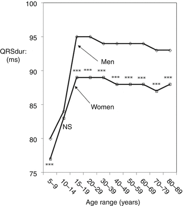
The evolution and the appearance of sex differences in QRS duration and heart rate was documented in a Canadian pediatric material [1] and confirmed in a larger normal study population by Mason et al. [2] covering a wide age range of children and adults (Table 2.1). QRS duration in age groups 15–19 and 20–29 years was 6 ms shorter and heart rate was 5 bpm faster in women than in men. Sex differences in QRS duration are further highlighted in Fig. 2.1.

Table 2.1
Heart rate and QRS duration in normal men and women by age
Sample size | Heart rate (bpm) | QRS duration (ms) | ||||
|---|---|---|---|---|---|---|
Women | Men | Women | Men | Women | Men | |
5–9 | 335 | 524 | 86 ± 13.5 | 82 ± 12.3 | 77 ± 9.5 | 80 ± 9.8 |
10–14 | 259 | 443 | 76 ± 11.9 | 75 ± 12.1NS† | 83 ± 9.5 | 84 ± 10NS |
15–19 | 310 | 333 | 70 ± 12.0 | 65 ± 11.0 | 89 ± 8.7 | 95 ± 8.8 |
20–29 | 2,469 | 2,528 | 69 ± 11.3 | 64 ± 11.7 | 89 ± 9.4 | 95 ± 9.3 |
30–39 | 3,954 | 3,411 | 70 ± 11.0 | 67 ± 11.7 | 89 ± 9.5 | 94 ± 9.4 |
40–49 | 7,633 | 5,362 | 70 ± 11.0 | 68 ± 11.6 | 88 ± 9.3 | 94 ± 9.4 |
50–59 | 6,340 | 5,344 | 69 ± 11.2 | 69 ± 12.0NS | 88 ± 9.6 | 94 ± 9.3 |
60–69 | 5,067 | 4,466 | 68 ± 11.4 | 68 ± 12.3NS | 88 ± 9.4 | 94 ± 9.6 |
70–79 | 3,745 | 2,901 | 67 ± 10.9 | 65 ± 11.6 | 87 ± 9.6 | 93 ± 9.7 |
80–89 | 1,291 | 864 | 67 ± 10.8 | 64 ± 11.0 | 88 ± 10.0 | 93 ± 9.6 |

Fig. 2.1
Evolution of QRS duration with age showing the appearance of gender differences during adolescence and prevailing through adulthood although gradually diminishing in magnitude with age (From Rautaharju et al. [4] with permission)
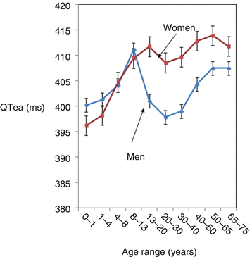
The rate-adjusted QT is commonly thought to be due to QT prolongation in women. A Canadian investigation on the age-evolution of QT reported in 1992 that gender difference in rate-adjusted QT arises from QT shortening in adolescent males rather than QT prolongation in females [3] (Fig. 2.2). The findings of the Canadian study were confirmed by a more recent study in a large group of women and men covering a wide age range [4]. The observed shorter QT in men than in women is unexpected because QRS duration is longer in men than in women reflecting higher left ventricular mass. More recent studies have demonstrated that the shorter rate-adjusted QT in men than in women is associated with an earlier start and earlier end of left ventricular (LV) epicardial repolarization [5, 6]. Data from Rautaharju et al. [7] in Table 3.2 in Chap. 3 will show that LV epicardial repolarization time (RTepi) is shorter in men than in women. QRS duration does not contribute to LV lateral wall RTepi or endocardial RT (RTendo) because of the reverse sequence of repolarization. However, QRS duration still contributes to QT prolongation during the terminal repolarization which is predominantly concordant with respect to depolarization sequence.


Fig. 2.2
Evolution of the rate-adjusted QTend interval with age showing the appearance of gender differences during adolescence persisting during adulthood although gradually diminishing in magnitude (From Rautaharju et al. [3])
A more detailed evaluation of sex differences in QT evolution in Chap. 9 shows that neurohormonal factors primarily testosterone appear as likely common mechanisms of gender differences in the evolution of ST elevation and QT.
2.2 Gender Differences in the Evolution with Age of ST J-Point Amplitude
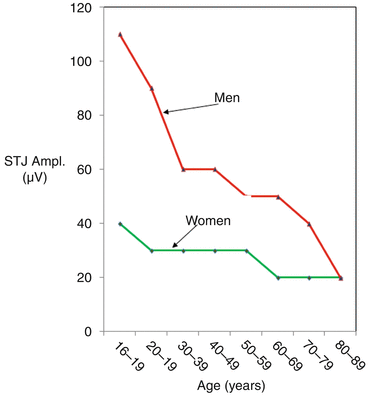
Ezaki et al. documented gender differences in the age-evolution of ST amplitudes in a study population of 640 subjects from 5 to 89 years in age [8]. These investigators observed a profound increase in STJ-point amplitude in men in age group 15–19 years with no increase in women in this age group or older. The sex difference decreased after age 30 years because ST amplitudes in men decreased gradually in men but ST segment amplitudes in men remained significantly higher in men than in women in all age groups of adults.
Rijnbeek et al. documented normal values derived from a large group (N = 13,354) of Dutch clinically normal men and women ranging in age from 19 to 90 years [9]. Figure 2.3 adapted from Supplementary Table 8 of that report shows the median values of the ST J-point amplitudes by age in women and men. The graphs show that J-point amplitude in adolescent males is distinctly higher than in females, confirming the observation of Ezaki et al. As in Ezaki’s report, the gender difference in J-point amplitude gradually levels off in older age groups.


Fig. 2.3
Age-evolution of the median values of ST J-point amplitudes in women and men from adolescence to old age (Adapted from Supplemental Table 8, Rijnbeek et al. [9])
2.3 Gender Differences in Waveforms
Gender differences between 529 men and 544 women 5–96 years in age were documented by Surawicz et al. [10] for ST waveforms in chest leads, with particular attention to V2 lead. Male and female patterns were defined by J-point amplitude and ST segment slope in a 60 ms window starting at J-point. STJ amplitude ≥0.1 mV with a slope ≥20° was used to define the male pattern. The female patterns was defined by STJ <0.1 mV and apparently a slope between 0° and 20°. ST patterns not meeting these criteria were defined as indeterminate. The proportion of female pattern was high in all age groups of women and changed relatively little with age (Table 2.2). The proportion of male patterns during adolescence increased in men and there was a profound decline in the proportion of male patterns in men starting in age group 25–35 years, apparently reflecting diminishing testosterone levels with age. Sex differences in proportions of male patterns in men and female patterns in women in the report of Surawicz et al. are further highlighted in Fig. 2.4.
Table 2.2
Distribution of male, female, and indeterminate ECG patterns among males and females of different age groups
Patterns in men (%) | Patterns in women (%) | |||||||
|---|---|---|---|---|---|---|---|---|
Age | No. | Male | Female | Indetemined | No. | Male | Female | Indetermined |
5–7 | 49 | 63.3 | 32.7 | 4.1 | 60
Stay updated, free articles. Join our Telegram channel
Full access? Get Clinical Tree
 Get Clinical Tree app for offline access

| |||