Fig. 18.1
24-h patterns in heart rate (HR) and systolic and diastolic BP (upper/lower BP curve) in ten male (closed) and ten female (open) healthy volunteers of an age of 21–23 years, motility was registered to control sleep activity. Data from [11] with permission
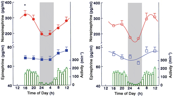
Concerning the plasma norepinephrine concentrations, however, there was a clear sex-dependent difference, the early morning rise in norepinephrine was significantly steeper in males than in females though both groups were normotensive (Fig. 18.2). In males, this observation could be an early sign of a genetically set greater early morning drive in sympathetic tone, which is often found in white coat hypertensive patients [12].


Fig. 18.2
24-h values in plasma norepinephrine and epinephrine concentrations and motility in healthy young (21–23 years) subjects. Figure from [11] with permission
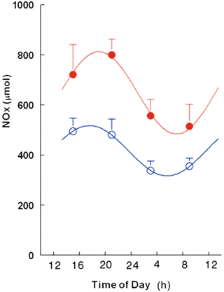
The sex-dependent difference was even greater for the NOx concentration determined in 6-h portions in the urine over 24 h in male and female subjects (Fig. 18.3). In both groups, NOx concentration displayed a 24-h rhythm with a peak value in the activity phase, similar to the rhythm in nitrate and cGMP secretion described by Bode-Boger et al. [10].


Fig. 18.3
Amount of NOx (nitrate/nitrite) in 6-h portions of urine in 10 female (filled) and 11 male (open) healthy volunteers. Note: NOx is significantly higher in females than males (p < 0.05). Figure from [11] with permission
These data clearly demonstrate that at an early age and under normotensive conditions, the important co-regulator of the vascular bed, NO, displays sex-dependent variations with higher values in female subjects, possibly allowing a better buffering capacity for BP changes for females than for males. This observation could contribute to the increasing data that show that the setting of the cardiovascular system is different between males and females. Though the therapeutic consequences are not clear at the moment, future clinical research must concentrate on cardiovascular factors associated with gender in more detail than has been the case in the past.
Data from Animal Experiments
Basic research in the cardiovascular system of experimental animals has been greatly improved by the availability of telemetric blood pressure monitoring devices. They are regarded as the gold standard today allowing in vivo experimentation in freely moving unrestrained animals [13–16], which is not the case for the tail-cuff method [17, 18]. Radiotelemetry also allows a closer look on the rhythmic organization of the cardiovascular system. These clinical experiments in small animals mainly used males. Thus, the data on cardiovascular effects and mechanisms in female rats are sparce. By 1993, we could demonstrate specific circadian rhythms in BP and HR in four different strains of rats, both normotensive and hypertensive [15]. In this study, there was evidence that in a transgenic rat strain (TGR), a dissociation occurred between BP and HR; whereas HR peaked in the activity phase of the animals at night, BP showed peak values in the resting phase during daytime [15], exhibiting a BP pattern known as non-dippers in humans. Thus, this rat strain displayed an internal desychronization concerning BP and HR.
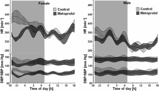
Maris et al. studied by radiotelemerty BP and HR in normotensive Wistar-Kyoto (WKY) and spontaneous hypertensive (SHR) rats [19]. In both strains and in males and females, they show significant daily variations, HR was higher in female WKY than in all other groups, and male SHR had higher systolic BP than the females [19]. Very similar to these findings, we describe that under control conditions of 12:12 h of light and darkness, the HR was higher in pro-estrous females than in age-mached male SHRs, whereas BP was inversely higher in males than in females [20] (Fig. 18.4). Urinary NO excretion was about threefold higher in female than in male SHR with peak value in the dark span [20]. Additionally, eNOS expression in the aortae of females was significantly higher than in males (relative expression 42.9 ± 7.3 vs. 25.2 ± 1.0, p < 0.05), indicating that elevated NOx excretion in females in comparison to males is linked to higher eNOS expression [20]. The beta-receptor blocking drug metoprolol was also more effective in decreasing HR and BP in females than in males. These animal data greatly support the notion that sex-dependent variations have to be taken into account as they contribute to the regulation and drug effectiveness within the cardiovascular system, both in animal and in humans.


Fig. 18.4
Heart rate, systolic and diastolic BP after intraperitoneal application of 30 mg/kg MET at 19.00 h to female (n = 7) and male SHR (n = 13). Shown are mean values ±95 % confidence intervals. The dark period is highlighted. Data from [20] with permission
< div class='tao-gold-member'>
Only gold members can continue reading. Log In or Register to continue
Stay updated, free articles. Join our Telegram channel

Full access? Get Clinical Tree


