Fig. 2.1
D- and L-pattern of ventricular topology
2.1.2 Description of the Right Ventricle
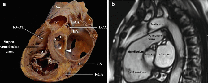
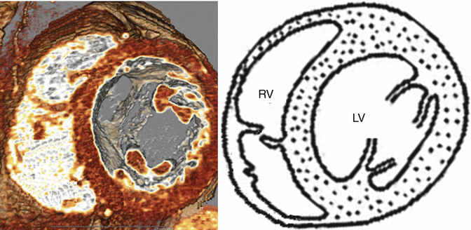
The right heart chambers are located anterior to their left counterparts. In general, the greater part of the anterior surface of the ventricular mass is occupied by the morphologically RV, which forms nearly the entire sternocostal surface of the heart and the inferior border of the cardiac silhouette (Fig. 2.2). The most apparent of the great vessels is the proximal pulmonary trunk together with its infundibulum when the heart is accessed from anterior. The proximate retrosternal location of the RV renders its echocardiographic approach more difficult and makes it more prone to injury during median sternotomy, especially when dilated or in redo operations. The shape of the RV is triangular from the side and is crescent shaped in cross section, having inferior diaphragmatic, anterior sternocostal, and posterior pulmonary surfaces. The margin between the first two surfaces is sharp and it is described as the acute margin. It is important to emphasize that the RV is an asymmetric chamber, which resembles a crescent shape, and it is wrapped around the right and anteroseptal side of the left ventricle (Fig. 2.3) [9]. Thickness of RV myocardium is normally less than 5 mm. Exceeding this limit is an indication of hypertrophy [11].


Fig. 2.2
Parasagittal section through the heart (a: human heart specimen; b: MRI image), illustrating the reciprocal relationships between the right and left chambers. The RV is most anterior and the right ventricular outflow winds around the aortic root (arrowhead). Ao aorta, CS coronary sinus, LAA left atrial appendage, LCA left coronary artery, PT pulmonary trunk, RCA right coronary artery, RVOT right ventricular outflow tract, R, L, and NF right, left and non-coronary aortic valve leaflets, A, S, and P tricuspid valve leaflets, anterior, septal, and posterior, respectively

Fig. 2.3
Computed tomographic image that clearly shows characteristic orientation of the crescent-shaped RV that is wrapped around the LV
The RV can be divided into either two or three components. From the two-compartment perspective, the RV consists of the sinus (the pumping chamber) and the infundibulum [2]. From the three-compartment point of view, the RV consists of inlet, apical trabecular, and outlet parts. This tripartite concept appears more correct from the embryological point of view and more useful, as one or more of the three components may be lacking in malformed hearts [12]. The inlet portion of the RV contains, and is limited by, the TV and its tension apparatus. The anatomy and pathophysiology of the inlet component are discussed later in this chapter together with the TV.
The interior of the RV appears heavily trabeculated. The apical trabecular part of the ventricle has particularly coarse trabeculations, which often form ridges along the inner surface of the wall or cross from one wall to the other. This represents the most constant feature of the RV in malformed hearts. The highly trabeculated RV present several muscle bands, including three most prominent bands: (i) the parietal band which along with the infundibular septum forms the crista supraventricularis, (ii) the septomarginal band which extends inferiorly and becomes continuous with the moderator band, and (iii) the moderator band, which crosses from the lower ventricular septum to the anterior wall, where it attaches to the anterior papillary muscle. It carries within a fascicle of the right bundle branch of the atrioventricular conduction system. The crista supraventricularis functionally integrates mechanical events during systole to narrow the orifice of the TV and to cause the RV free wall to move toward the septum propelling blood into the lungs. There are three papillary muscles in the RV: the anterior, posterior, and septal. The anterior (attached to the anterior wall of the RV) is large, with chordae inserted to the anterior and posterior cusps of the TV. The posterior (attached to the inferior wall of the RV) is often represented by two or more parts, with chordae inserted to the posterior and septal cusps, as well as a variable group of small septal papillary muscles (attached to the interventricular septum), with chordae inserted to the anterior and septal cusps [13].
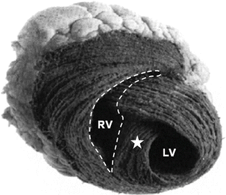
The myocyte arrangement is two layered in RV wall and differs from that of the three-layered LV. The fibers of the superficial layer of the RV are arranged circumferentially in a direction that is parallel to the atrioventricular groove in continuity with the LV. The deep musculature of the RV is longitudinally aligned base to apex (in contrast to the LV, where oblique fibers are found superficially, longitudinal ones on the endocardium and circumferential in between) [14]. Hence, there is a different contraction pattern between the inflow and outflow tracts of the RV [15, 16] as well as different timing in contraction; the RV inflow region contracts before that of the outflow, resulting in a peristaltic action [17]. Moreover, the two main parts of the RV contract perpendicular to each other: the inflow component longitudinally and the outflow circumferentially. This is due to the fact that the inflow portion of the RV is mainly composed of circumferential fibers, while the outflow portion is composed mainly of fibers running longitudinally [18] (Fig. 2.4). This pattern explains differential response to inotropes. The inotropic response of the outflow tract is greater than that of the inflow tract, probably as a mechanism to protect the pulmonary vasculature from high pressure [20].


Fig. 2.4
Structure of the RV. Note how the RV free wall wraps around the LV creating a crescent-shaped cavity, while the LV is conical in shape. The interventricular septum (star) is a single structure that consists of fibers which also form the free wall of the LV. This special architecture determines interaction between the two ventricles, which is described as ventricular interdependence (see Chap. 3) (Adapted from Saleh et al. [19])
The blood ejection pattern from the RV can be summarized as follows: (i) longitudinal shortening (the tricuspid valve annulus moves in the apical direction (currently determined by measuring TAPSE, i.e., tricuspid annular plane systolic excursion)), (ii) the compression of the RV chamber by contraction of the transverse fibers (the bellows action), (iii) traction on the free wall of the RV, and (iv) the contraction of the interventricular septum and the “wringing” action of the LV; therefore, it becomes evident that LV function remains an important determinant of RV ejection [21].
2.1.3 Blood Supply
The RV is perfused by both coronary arteries, mainly the right coronary artery (RCA), which marks the right border of the ventricle. While the infundibulum and anterior RV wall receive a more constant arterial supply (conal artery), the vascularization of the inferior/diaphragmatic wall and posterior septum depends on the coronary dominance [2]. Arterial branches from the left coronary system supply the free wall of the RV adjacent to the anterior part of the septum, the apical area of the RV, while the RCA is almost always the main arterial supply toward the base of the RV. Moreover, the RCA perfuses the sinus and the atrioventricular nodes (the latter just in cases of right dominant perfusion). The right marginal arteries supply blood to the lateral wall of the RV. Blood supply from the RCA to the RV is about equal during systole and diastole, in contrast to perfusion pattern of the LV which is mostly during diastole [22].
As analyzed in physiology chapter, the RV appears relatively resistant to ischemia. This is related to many factors: extensive collateral circulation (the moderator band artery of the RV originates from the first septal branch of the left anterior descending artery), numerous thebesian vessels that the RV possesses which offer alternative pathways, and higher systolic/diastolic coronary blood flow ratio, reduced oxygen demand, and increased oxygen extraction capacity [22, 23].
2.2 Differences Between Right and Left Ventricles
The right and left ventricles are anatomically, physiologically, and functionally distinct, although there is continuity between the muscle fibers, which functionally binds the two ventricles together. The morphological differences between the left and right ventricles are (i) a more apically situated hinge point of the septal leaflet of the tricuspid valve relative to the anterior leaflet of the mitral valve, (ii) the presence of a moderator band in the RV cavity, (iii) more than two papillary muscles, (iv) a trileaflet atrioventricular valve with septal attachments, (v) predominantly coarse trabeculations, and (vi) a ventriculo-infundibular fold that separates the tricuspid valve from the pulmonary valve (as opposed to the aortomitral continuity of the LV) [9]. It should be noted that, for the cardiac morphologist, it is the uniformly coarse apical trabeculations that serve as the most constant anatomical feature of the morphologically RV [24].
The two ventricles appear totally different in shape: the LV can be described as almost cylindrical with a conical apex, while the RV pyramidal or crescent shaped. The wall is about three times as thick in the LV (7–11 mm vs. 2–5 mm). Thus, the muscle mass of the RV is approximately one-sixth that of the LV (17–34 g/m2 vs. 65–110 g/m2, respectively). On the contrary, RV end-diastolic volume is slightly greater rather than that of the LV (49–101 ml/m2 vs. 44–89 ml/m2). This renders it capable of accommodating increased preload, in contrast to limited adjustment to increased afterload.
2.3 Right Atrium
The right atrium (RA) has triangular shape and comprises four basic parts, (i) the venous component receiving the systemic venous return, (ii) the appendage, (iii) the (small) body, and (iv) the vestibule of the tricuspid valve [24]. It is separated from the left atrium with the septum.
The venous components of the two chambers are derived from different embryonic sources, with the entirety of the embryonic systemic venous sinus being incorporated into the morphologically RA, in contrast to the pulmonary venous component which is derived from mediastinal myocardium, as are the components of the atrial septum [25]. The anatomical criteria of distinguishing morphological right from left atrium are mainly connections of caval veins and identification of limb of the oval fossa [26]. The superior vena cava (SVC) enters the atrium in the right anterior portion of the superior wall, while the inferior vena cava (IVC) enters into the right posterior portion of the inferior wall. The mouth of the IVC is looking toward the floor of the oval fossa, which has an implication in fetal life directing the blood toward the left atrium. The diameter of the IVC represents an important echocardiographic diagnostic tool as presented in echocardiography chapter. The coronary sinus (CS) collects all the venous return from the heart and empties into the infero-posterior portion of the right atrium, just above the tricuspid annulus.
Morphologically, the posterior smooth wall of the right atrium, which measures almost 2 mm in thickness, and the anterior trabeculated appendage are marked by a well-formed muscle bundle, the crista terminalis, which is identified externally by the prominent terminal groove. This is reinforced in the fetal life by sheetlike structures, which separate the orifices of the IVC from the atrial appendage. These become the valves of the IVC (eustachian valve) and the CS (thebesian valve) and may be seen to variable extent in the adult heart. A tendinous structure extends intramyocardially through the sinus septum in most hearts, being a continuation of the commissure between the eustachian and thebesian valves. This is called the tendon of Todaro and is a vital structure as it demarcates the position of the atrioventricular node. The vestibule to the RV surrounds the orifice of the TV and is contiguous with both the systemic venous component and the appendage of the RA.
When inspecting the right atrium through a surgical incision along the terminal groove, we observe an extensive septal surface between the openings of the caval veins and the orifice of the TV [24]. The apparent extent of this septum is considered “spurious” by RH Anderson [27]; the “true” septum is confined to the floor and the anteroinferior margin of the oval fossa, while the superior rim of the oval fossa is produced by the folds of the interatrial groove. This separates the orifice of SVC and the entrance of the pulmonary veins to the left atrium [28]. The posteroinferior rim is formed by reflection of the musculature forming the opening of the CS and the orifice of the IVC. These muscular structures continue anteriorly within the atrium as the eustachian ridge.
The anatomy of RA does not relate directly to the pathophysiology of right heart failure. It is the involvement of the RA to the conduction system and its elements, which makes it important in heart failure. In the vast majority (90 %) of the human population, the sinus node is positioned inferiorly within the terminal groove relative to the crest of the appendage. In the remaining, the node lies in a horseshoe fashion across the crest of the appendage [29]. Arterial supply to the sinus node is also of clinical significance. The sinoatrial artery emerges from the proximal segment of the RCA in about 55 % of individuals and from the circumflex artery in the remainder [24]. It courses through the anterior interatrial groove toward the superior cavoatrial junction running within the atrial myocardium. A more lateral origin is found in patients with congenital malformations [30]. This abnormal course should be taken into account when planning atrial surgical incisions. The atrioventricular node is contained within the well-known “triangle of Koch.” This important landmark is bounded by the tendon of Todaro, the attachment of the septal leaflet of the tricuspid valve, and the orifice of the CS. The main pathways for conduction toward the atrioventricular node are the terminal crest and the margins of the oval fossa [31]. Bachmann’s bundle is responsible for conduction to the left atrium. Additional pathways exist through the eustachian ridge and the CS [32]. Abnormal atrial rhythms can be initiated at several sites along the terminal crest, where areas of conducting (primary) cardiomyocytes are joined with working myocardium. In order to avoid postoperative atrial arrhythmias, meticulous care should be given on preserving the sinus and atrioventricular nodes and their arteries, rather than concern about nonexistent tracts of purportedly specialized atrial myocardium.
Stay updated, free articles. Join our Telegram channel

Full access? Get Clinical Tree


