Fig. 2.1
Four-chamber view of the fetal heart. LV left ventricle, RV right ventricle, RA right atrium. ★ denotes marks enlarged coronary sinus giving false impression of a primum septal defect

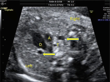
Fig. 2.2
Fetal three-vessel and tracheal view. Left and right SVC denoted by arrows. A aorta, D duct, T trachea
Discussion
Optimal fetal cardiac imaging depends on a combination of factors, including fetal lie and maternal habitus. Whilst in some patients it is possible to get pictures of comparable quality to a postnatal echo, in others this can be challenging. It is in these situations we have to be particularly wary of common pitfalls in diagnosis.
The coronary sinus lies behind the left atrium and becomes enlarged when it receives additional flow. The most common cause of additional flow is drainage of a left SVC to the coronary sinus that occurs in approximately 0.3 % of the population. More rarely the coronary sinus may appear enlarged due to anomalous pulmonary venous drainage.
< div class='tao-gold-member'>
Only gold members can continue reading. Log In or Register to continue
Stay updated, free articles. Join our Telegram channel

Full access? Get Clinical Tree


