(1)
Department of Neuroscience, University of Turin Ospedale Molinette, Turin, Italy
Abstract
These are represented by the superior and inferior ophthalmic veins, which drain posteriorly in the cavernous sinus. They communicate anteriorly with the facial and frontal veins. There are connections with the pterygoid plexus (see also “Superior– Inferior Ophthalmic Vein” in Chap. 9) (Fig. 10.1).
10.1 Orbital Veins
The orbital veins develop from the primitive maxillary and supraorbital veins, tributaries of the anterior part of the primitive dural plexus or primary head sinus (see also Sect. 10.9). The result of this embryological evolution are the superior and inferior ophthalmic veins, which drain posteriorly in the cavernous sinus. They communicate anteriorly with the facial and frontal veins. There are connections with the pterygoid plexus (see also “Superior– Inferior Ophthalmic Vein” in Chap. 9) (Fig. 10.1).
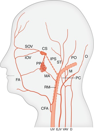
Fig. 10.1
Internal jugular vein (IJV), retromandibular vein (RM), to which converge the superficial temporal vein (ST) and the maxillary vein (MA). Pterygoid plexus (PP), facial vein (FA), common facial vein (CFA), formed by the confluence of the RM and FA entering the IJV. Posterior auricular vein (PO), superficial temporal vein (ST), occipital vein (O), external jugular vein (EJV), to which converge PO and O. Mastoid vein (M), posterior condylar vein (PC), deep cervical vein (D), to which drain partially the occipital vein and venous plexus of the vertebral artery (VAV), with some of its connections. Superior–inferior ophthalmic veins (SOV–IOV), connected anteriorly with the facial and frontal veins and posteriorly with the cavernous sinus (CS). Inferior petrosal sinus (IPS)
10.2 Facial Veins
The anterior facial vein begins in the naso-orbital angle, with the angular vein, which has an anastomosis with the ophthalmic veins. The anterior facial vein runs inferiorly and posteriorly in an oblique course along the face; it crosses over the mandible, and shortly after it is joined by the retromandibular vein; together they form the common facial vein, which enters the internal jugular vein at the level of the hyoid bone (Osborn 1999).
The anterior facial vein receives tributaries from the orbit, lips, and facial skin and muscles and from the menton–submental region. Occasionally, the lingual and superior thyroid veins also join the anterior facial vein, instead to drain separately into the internal jugular vein. The anterior facial vein is connected posteriorly with the pterygoid venous plexus through the deep facial vein.
10.3 Retromandibular Vein
Also known as the posterior facial vein, the retromandibular vein arises at the junction of the maxillary vein and superficial temporal vein, below the neck of the mandible. The maxillary vein drains the pterygoid venous plexus. This latter is a venous network lying between the pterygoid muscles, which receives numerous tributaries from the naso-oral and masticator areas. The pterygoid venous plexus is connected with the anterior facial vein through the deep facial vein and intracranially with the paracavernous–cavernous sinus. The superficial temporal vein drains the temporal region.
Stay updated, free articles. Join our Telegram channel

Full access? Get Clinical Tree


