Fig. 9.1
A representative of an apical suction heart positioner: X-pose® (MAQUE) and other devices for off-pump coronary artery bypass grafting (MAQUE)
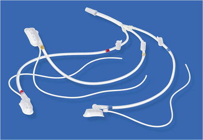
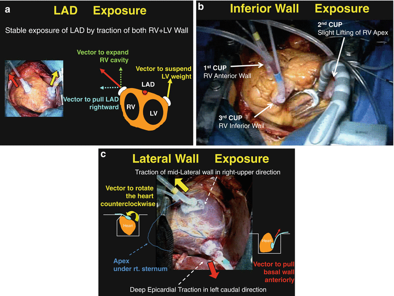
We developed a multi-suction cardiac positioner, TENTACLES™ (Sumitomo Bakelite Co., Ltd, Tokyo, Japan) (Fig. 9.2) [8]. This device has three independent small suction cups and arms with elastic silicone strings. Due to the high tissue affinity of the suction cup, the suction cups can be applied on any surface of the heart and pulled in any desired three different directions using traction string, which is fixed to the surgical drape using clamp. To visualize the LAD, we typically apply one suction cup on the anterior wall of the RV and pull the RV rightward. To secure the heart position, we usually apply another additional suction cup at the LV anterior wall and fix it loosely to the left side (Fig. 9.3a). But anastomosis to the LAD can be performed even with only one or two suction cups applied on the RV, owing to the powerful suctioning capacity of this device. In case of unstable heart such as acute anterior wall myocardial infarction or low ejection fraction, this LV non-touch maneuver is especially useful to avoid suction-induced injury to the weakened infarcted myocardium, hypotension, or life-threatening arrhythmia induced by touching the LV wall. To visualize the inferior wall, we usually apply one suction cup on the RV anterior wall and pull it cranially and another cup on the inferior wall near the apex and slightly lift the apex under the sternum. In addition, we apply third suction cup on the RV inferior wall near the acute margin and pull it in the right-cranial direction to expand exposure of the inferior wall (Fig. 9.3b). The PD can be easily exposed under stable hemodynamic conditions. To visualize the lateral wall, we apply the first suction cup on the apex and lift the heart to confirm the target coronary artery. The second suction cup is applied on the LV lateral wall to rotate the heart rightward, and the traction string is fixed at the right side drape. The third suction cup is applied deeply near the crux of the heart and pulled in the left-caudal direction, and then fixed at the left side drape. Heart is rotated toward the right chest cavity (Fig. 9.3c). Finally, the first suction cup on the apex is detached. The apex is displaced under the right-sided sternum. Additional application of one suction cup on RV outflow tract may sometimes be helpful to prevent RV outflow kinking.


Fig. 9.2
A multi-suction heart positioner: TENTACLES™ (Sumitomo Bakelite Co., Ltd, Tokyo, Japan)

Fig. 9.3
Exposure of coronary arteries using TENTACLES™. (a) Exposure of left anterior descending artery. (b) Exposure of posterior descending artery. (c) Exposure of posterior lateral artery
According to the annual report of Japanese Association for Coronary Artery Surgery 2012, suction heart positioner was utilized in 87 % of overall Japanese coronary surgery units.
9.4 Hemodynamic Change Caused by Heart Displacement
Hemodynamic changes are observed in any heart displacement technique. Although both deep pericardial traction and vacuum-assisted suction are safe and effective maneuvers to expose the coronary artery in OPCAB, vacuum-assisted suction devices appear to produce lesser hemodynamic impairment than the deep pericardial traction technique [9, 10]. Transesophageal echocardiography is very useful for quickly detecting signs of hemodynamic instability [11]. Hemodynamic changes are considered to result from RV kinking and obstruction, regional wall motion abnormalities caused by transient ischemia, or compression of the heart by stabilizers. Mitral annulus distortion by heart displacement can cause mitral regurgitation [12].
Stay updated, free articles. Join our Telegram channel

Full access? Get Clinical Tree


