Fig. 3.1
Bypass site and graft arrangement
Overall bypass occlusion rate was 3.1 % (366/11,968). Graft stenosis rate was 1.9 %. Overall bypass occlusion rate was 2.1 % in LITA, 1.9 % in RITA, 3.3 % in RA, 6.3 % in SVG, and 4.2 % in GEA. There were 297 patients with at least one occluded bypass. Univariate analysis showed that the risk factors of patients with occluded bypass grafting were female gender, weight, height, diabetes, blood creatinine level, liver dysfunction, non-left main trunk lesion, three-vessel disease, family history of ischemic heart disease, more than four distal anastomoses, and no use of preoperative β blocker and aspirin (Table 3.1). OPCAB or prior percutaneous coronary intervention was not a risk factor. In multivariate analysis, only female gender (HR, 1.53: 95 % CI, 1.13–2.07), liver dysfunction (HR, 2.09: 95 % CI, 1.18 to 3.70), non-left main trunk lesion (HR, 1.54: 95 % CI, 1.14–2.08), family history of ischemic heart disease (HR, 1.52: 95 % CI, 1.07 to 2.17), and more than four distal anastomoses (HR, 1.34: 95 % CI, 1.01–1.77) were risk factors (Table 3.2). Bypass occlusion was related to operative morbidity (p = 0.003) and all morbidity (p = 0.01). Bypass occlusion was not related to operative death or hospital death (Table 3.3).
Table 3.1
Patient characteristics in relation to graft occlusion
Univariate analysis | |||
|---|---|---|---|
Variable | With all patent grafts (n = 3235) | With occluded graft (n = 297) | p value |
Age (year) | 65.5 ± 1.1 | 66.5 ± 9.6 | 0.13 |
Female | 694 (21 %) | 84 (28 %) | 0.01 |
Weight (kg) | 61.2 ± 10.6 | 59.6 ± 11.3 | 0.015 |
Height (cm) | 160.2 ± 8.9 | 158.0 ± 11.0 | 0.0002 |
BMI (kg/m2) | 23.9 ± 5.3 | 24.1 ± 8.3 | 0.58 |
Hypertension | 2149 (66 %) | 187 (63 %) | 0.29 |
Hypercholesterolemia | 1741 (54 %) | 157 (53 %) | 0.97 |
Diabetes | 1659 (51 %) | 131 (44 %) | 0.02 |
Oral medication | 445 (14 %) | 50 (17 %) | 0.006 |
Insulin | 452 (14 %) | 35 (12 %) | 0.83 |
Creatinine (mg/dl) | 1.67 ± 2.22 | 2.45 ± 1.00 | 0.04 |
Dialysis | 208 (6 %) | 17 (6 %) | 0.97 |
Liver dysfunction | 97 (3 %) | 18 (6 %) | 0.009 |
CTR by chest X-ray | 50.6 ± 5.6 | 50.8 ± 5.2 | 0.64 |
NYHA class III, IV | 578 (18 %) | 56 (19 %) | 0.22 |
CCS class III, IV | 710 (22 %) | 69 (23 %) | 0.14 |
Prior PCI | 1354 (42 %) | 102 (34 %) | 0.06 |
Prior MI | 1107 (34 %) | 98 (33 %) | 0.59 |
LVEF | 55.6 ± 14.8 | 57.3 ± 14.4 | 0.12 |
EF <0.35 | 195 (6 %) | 16 (5 %) | 0.21 |
Non-LMT lesion | 1981 (61 %) | 205 (69 %) | 0.01 |
Three-vessel disease | 2190 (68 %) | 225 (76 %) | 0.005 |
Family history of IHD | 397 (12 %) | 46 (15 %) | 0.03 |
Emergent surgery | 215 (7 %) | 16 (5 %) | 0.40 |
OPCAB | 2754 (85 %) | 261 (88 %) | 0.20 |
≥4 distal anastomoses | 1474 (46 %) | 157 (53 %) | 0.02 |
β blocker | 744 (23 %) | 67 (23 %) | 0.047 |
Aspirin | 1789 (55 %) | 123 (41 %) | 0.14 |
Table 3.2
Patient characteristics in relation to graft occlusion
Multivariate analysis | |||
|---|---|---|---|
Variable | p value | Hazard ratio | 95 % CI |
Female | 0.007 | 1.53 | 1.13–2.07 |
Liver dysfunction | 0.01 | 2.09 | 1.18–3.70 |
Non-LMT lesion | 0.005 | 1.54 | 1.14–2.08 |
Family history of IHD | 0.02 | 1.52 | 1.07–2.17 |
≥4 distal anastomoses | 0.04 | 1.34 | 1.01–1.77 |
Table 3.3
Outcomes in relation to graft occlusion
Outcomes | With all patent grafts (n = 3235) | With occluded graft (n = 297) | p value |
|---|---|---|---|
All morbidity | 932 (29 %) | 106 (36 %) | 0.01 |
Operativea | 105 (3 %) | 20 (7 %) | 0.003 |
Infection | 182 (6 %) | 22 (7 %) | 0.24 |
Neurological | 59 (2 %) | 4 (1 %) | 0.57 |
Pulmonary | 105 (3 %) | 15 (5 %) | 0.12 |
Renal | 97 (3 %) | 10 (3 %) | 0.69 |
Othersb | 590 (18 %) | 65 (22 %) | 0.19 |
Operative death | 17 (0.5 %) | 1 (0.3 %) | 0.98 |
Hospital death | 44 (1.4 %) | 2 (0.7 %) | 0.33 |
Graft occlusion rate in relation to graft material and cardiopulmonary bypass was shown in Fig. 3.2. The occlusion rate of SVG (7.3 %) was significantly (p < 0.001) higher than LITA (1.7 %), RITA (1.7 %), RA (3.4 %), and GEA (3.8 %) in OPCAB. The occlusion rate of RA and GEA was also significantly (p < 0.001) higher than LITA and RITA in OPCAB. In standard CABG patients, bypass occlusion rate was not statistically different between graft materials. The occlusion rate of RITA (1.7 %) in OPCAB was significantly (p = 0.046) lower than that (3.5 %) in standard CABG. On the contrary, the occlusion rate of SVG (7.3 %) in OPCAB was significantly (p = 0.032) higher than that (2.8 %) in standard CABG. As a whole there was no difference in the bypass occlusion rate between OPCAB (3.1 %) and standard CABG (2.9 %).
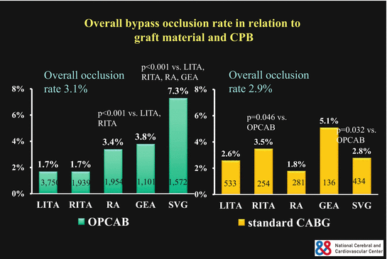

Fig. 3.2
Bypass occlusion rate
Bypass occlusion and stenosis was examined in relation to graft material and bypass area. Bypass occlusion and stenosis rate of RITA to left anterior descending artery (LAD) (6.1 %) was significantly (p = 0.013) higher than that of LITA to LAD (3.8 %) though the difference of occlusion rate did not reach statistical significance (p = 0.053) (Fig. 3.3). In the circumflex area bypass occlusion rate of SVG (7.1 %) was significantly higher than that of all arterial grafts of LITA (4.2 %, p = 0.030), RITA (1.7 %, p < 0.001), RA (2.2 %, p < 0.001), and GEA (3.7 %, p = 0.029). The occlusion rate of RITA was significantly lower than that of LITA (p = 0.002) and GEA (p = 0.046) in this area (Fig. 3.4). The occlusion rate of RA was significantly lower than that of LITA (p = 0.024) in the circumflex area. Bypass occlusion rate in the right coronary territory was not related to graft material (Fig. 3.5). However, overall bypass occlusion rate in the right coronary area (4.8 %) was significantly (p < 0.001, p = 0.015) higher than that in LAD (1.8 %) and circumflex area (3.5 %).
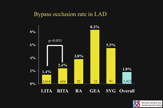
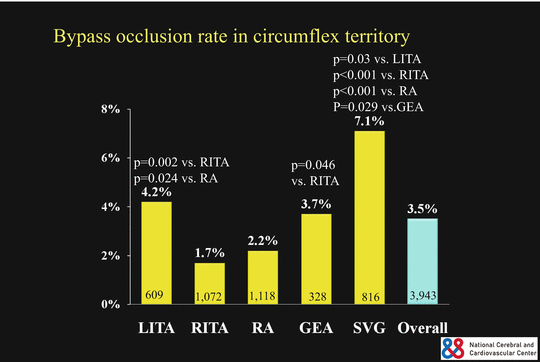
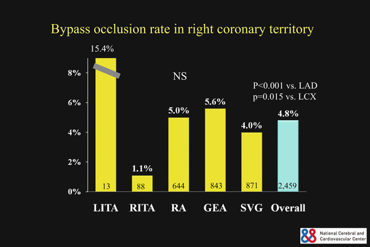

Fig. 3.3
Bypass occlusion rate in the LAD area

Fig. 3.4
Bypass occlusion rate in the circumflex area

Fig. 3.5
Bypass occlusion rate in the right coronary area
Bypass occlusion and stenosis in relation to the site of the right coronary artery (RCA) was examined. Bypass occlusion and stenosis of the RITA to the right main coronary artery (11.1 %) was significantly higher than that of RA (0 %, p = 0.004) and SVG (2.0 %, p = 0.013) (Fig. 3.6). Bypass occlusion and stenosis of the GEA to the main RCA was significantly higher (13.3 %) than that of RA (p < 0.001) and SVG (p < 0.001). Bypass occlusion and stenosis rate of GEA to the main right coronary artery was significantly (p = 0.009) higher than that to the posterior descending artery (5.1 %). Bypass occlusion and stenosis rate of SVG to the main RCA was significantly lower than that to the posterior descending artery (7.4 %, p = 0.045) and atrioventricular branch (11.6 %, p < 0.001).
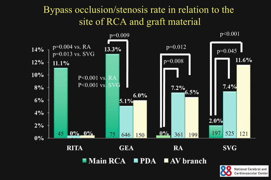

Fig. 3.6
Bypass occlusion and stenosis rate in relation to the site of right coronary artery
Bypass occlusion of the RA in relation to composite and aortocoronary bypass configuration was examined. The occlusion rate of RA as a composite graft was significantly (p = 0.0023) higher in the right coronary territory (5.4 %) than that in the circumflex territory (2.1 %) (Fig. 3.7). There was no significant difference in RA occlusion rate between composite and aortocoronary bypass both in the circumflex and the right coronary area.
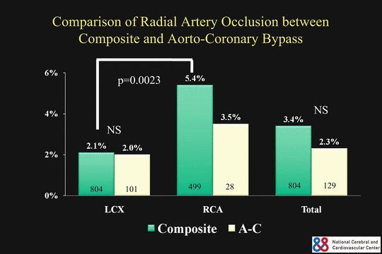

Fig. 3.7
Bypass occlusion rate of the radial artery in relation to composite and aortocoronary configuration
Female gender, liver dysfunction, non-left main trunk lesion, family history of ischemic heart disease, and more than four distal anastomoses were independent risk factors in patients who had at least one bypass occlusion. Women have more severe coronary disease, higher age, higher comorbidity, lower body surface area, and lower percentage of LITA use. However, even after the risk matching of these factors, it was confirmed that female gender is an independent risk factor for adverse events and mortality after CABG [30]. Smaller body surface area and fragile coronary artery were the major causes of higher risk of perioperative mortality and morbidity in women [31]. Early occlusion of the bypass graft may explain the higher mortality and morbidity in women according to our results. There was no previous report regarding liver dysfunction and bypass graft patency to my knowledge. Liver dysfunction or cirrhosis may cause coagulation abnormality after CABG. Risk factors of non-left main trunk lesion, family history of ischemic heart disease, and more than four distal anastomoses may be related to the severity and diffuseness of the coronary lesion. The difficulty and many graft anastomosis sites may be attributed to bypass occlusion.
In the present study, there were several interesting results regarding the graft material and the method of its use both in OPCAB and standard CABG. First, LITA was the best graft for LAD. The RITA was not as good as the LITA as a graft for LAD. As the multivariate analysis showed that the female gender was an independent risk factor of graft occlusion, distal RITA in women might be not appropriate for LAD grafting. Second, in the circumflex area, graft patency of RITA and RA were better than LITA, GEA, and SVG. There was no difference in the graft patency of RA and RITA in this area. RITA was used as a composite graft with LITA rather than in situ graft. There was no difference in the RA graft patency in this area between the method of composite and aortocoronary bypass in the present study. RA could be used as a composite graft as well as aortocoronary bypass in some previous studies [32, 33]. However, Gaudino and associates found flow competition more frequently in the composite RA conduits than in the aortocoronary RA conduit [34]. RA is suitable for the second arterial graft next to LITA as composite graft at least in the circumflex area. Third, in the right coronary territory, the overall graft patency was worse than LAD and circumflex area. Bypass occlusion rate in the right coronary territory was not related to graft material. However, RITA and GEA were not suitable for the graft to the main RCA probably because the coronary arterial wall was much thicker than the wall of RITA and GEA, which might cause graft stenosis and occlusion. For the main RCA RA and SVG grafts were better than the RITA and the GEA as in situ graft. RA graft patency in this site was not better than SVG. Considering the higher occlusion rate of the composite RA in this area compared to aortocoronary RA grafting, the RA as aortocoronary bypass to the right coronary branches might be favorable when total arterial grafting is aimed.
Total arterial coronary revascularization could avoid the problems associated with vein graft failure [35, 36]. Bilateral ITAs are the conduits of first choice because of excellent short- and long-term patency and improved survival [37, 38]. However, bilateral ITA harvesting has shown a higher incidence of sternal wound infection in patients taking insulin or steroids, who are obese or have chronic obstructive lung disease [30]. RITA to LAD graft crossing the midline of the chest may obstruct future reoperation for aortic valve surgery, but to avoid this situation, the RITA is unable to reach the left ventricular posterolateral wall if it passes through the transverse sinus. As the second arterial graft in addition to LITA to LAD anastomosis, some studies comparing RITA and RA showed the same clinical and angiographic results [39, 40].
The comparative performance of RA versus SVG is of considerable interest. Previous angiographic observational studies have shown that the RA achieved excellent short- (96–100 %), mid- (94–97 %), and long-term (84–96 %) patency [41]. Patency rates of the RA have exceeded those of the SVGs at all-time points. Only the Cleveland Clinic reported worse graft patency of the RA than the SVG [42]. Randomized comparison of midterm graft patency between the RA and the SVG at 5 years showed disappointing graft patency of RA (87 %) compared to SVG (94 %) in RAPCO trial [43]. However, these results were based on the small angiographic studies. The RSVP randomized trial was designed to compare 5-year patency rates of aortocoronary RA and SVG to the circumflex coronary artery. The graft patency of the RA (98.3 %) was significantly better than that of the SVG (86.4 %) [44]. In the RAPS trial, the RA was randomly assigned to bypass the major artery in either the right coronary territory or the circumflex coronary territory, with the SVG used for the opposite territory, which had proximal lesions at least 70 % diameter narrowing [45]. The graft occlusion of the RA (8.2 %) was significantly lower than that of the SVG (13.6 %) at 1 year. Diffuse narrowing of the graft was present in 7.0 % of the RA grafts and only 0.9 % of SVGs. The absence of severe native vessel stenosis was a risk of graft occlusion and diffuses narrowing of the RA. Patency of the RA grafts was similar in RCA and circumflex arteries. In our study graft occlusion rate of the RA (2.2 %) was better than that of the SVG (7.1 %) in the circumflex area, but the same in the right coronary territory (RA: 5.0 %, SVG: 5.6 %). In our early postoperative study, composite RA grafts showed competitive flow in the setting of <75 % proximal coronary stenosis especially in the right coronary branches [46].
Graft patency of RITA and GEA was lower in standard CABG than in OPCAB in this study. The reason could be explained as follows. In OPCAB cases grafting to the main RCA might be avoided because hypotension and bradycardia occur frequently when the native coronary stenosis is moderate. The main RCA is sometimes very thick and calcified compared to other territory of right coronary branches, which are not suitable for OPCAB. Bypass occlusion rate of RITA and GEA was higher when the target area was the main RCA compared to grafting to the right coronary branches.
In summary, the LITA was the best graft for LAD, and the RA was as good as the RITA and better than SVG in the circumflex area. SVG should be avoided in OPCAB due to postoperative hypercoagulability status. Graft patency in the RCA area was not related to graft material, and the RITA and GEA should be avoided as a graft to the main RCA.
3.5 Critical Appraisal of Recent RCT in Western Countries
The operative mortality, graft patency, and long-term outcomes in OPCAB compared with conventional CABG are still controversial. The 30-day mortality after primary elective CABG with CPB was less than 1.0 % in Japan in 2011 [2]. Therefore, it is not surprising that the recent RCTs in western countries showed no difference in operative mortality between OPCAB and on-pump CABG. Puskas and associates demonstrated that OPCAB using STS database disproportionally benefits high-risk patients and female gender [47, 48]. In elective CABG cases in Japan, the operative mortality of OPCAB and standard CABG has been similar since 2008 (Table 3.4). On the contrary, in all CABG cases, the operative mortality of standard CABG was twice as high as that of OPCAB last 10 years. In the real world OPCAB has decreased the mortality in high-risk patients at least in Japan. The operative mortality in conversion cases from OPCAB to CABG with CPB has been between 2.7 and 6.7 % even in primary elective CABG. Including all cases that were converted to CABG with CPB, the operative mortality was between 4.4 and 8.3 %. Cardiac center and cardiac surgeons should be accustomed to OPCAB as a daily operation from this data, although less than 10 % of patients who have CABG really need OPCAB. Otherwise unexpected conversion from OPCAB to CABG with CPB caused deleterious outcomes.
Table 3.4


Operative mortality of isolated CABG in Japan
Stay updated, free articles. Join our Telegram channel

Full access? Get Clinical Tree


