Evaluation of the Patient with Pulmonary Disease

3


Evaluation of the Patient with Pulmonary Disease


In evaluating the patient with pulmonary disease, the physician is concerned with three levels of evaluation: macroscopic, microscopic, and functional. The methods for assessing each of these levels range from simple and readily available studies to highly sophisticated and elaborate techniques requiring state-of-the-art technology.


Each level is considered here, with an emphasis on the basic principles and utility of the studies. Subsequent chapters repeatedly refer to these methods because they form the backbone of the physician’s approach to the patient.


Evaluation on a Macroscopic Level


Physical Examination


The most accessible method for evaluating the patient with respiratory disease is the physical examination, which requires only a stethoscope; the eyes, ears, and hands of the examiner; and the examiner’s skill in eliciting and recognizing abnormal findings. Because the purpose of this discussion is not to elaborate the details of a chest examination but to examine a few of the basic principles, the primary focus is on selected aspects of the examination and what is known about mechanisms that produce abnormalities.


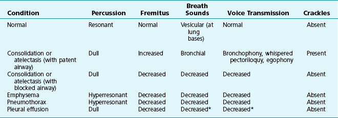
Apart from general observation of the patient, precise measurement of the patient’s respiratory rate, and interpretation of the patient’s pattern of and difficulty with breathing, the examiner relies primarily on palpation and percussion of the chest and auscultation with a stethoscope. Palpation is useful for comparing the expansion of the two sides of the chest. The examiner can determine whether the two lungs are expanding symmetrically or if some process is affecting aeration much more on one side than on the other. Palpation of the chest wall is also useful for feeling the vibrations created by spoken sounds. When the examiner places a hand over an area of lung, vibration normally should be felt as the sound is transmitted to the chest wall. This vibration is called vocal or tactile fremitus. Some disease processes improve transmission of sound and augment the intensity of the vibration. Other conditions diminish transmission of sound and reduce the intensity of the vibration or eliminate it altogether. Elaboration of this concept of sound transmission and its relation to specific conditions is provided in the discussion of chest auscultation.


When percussing the chest, the examiner notes the quality of sound produced by tapping a finger of one hand against a finger of the opposite hand pressed closely to the patient’s chest wall. The principle is similar to that of tapping a surface and judging whether what is underneath is solid or hollow. Normally percussion of the chest wall overlying air-containing lung gives a resonant sound, whereas percussion over a solid organ such as the liver produces a dull sound. This contrast allows the examiner to detect areas with something other than air-containing lung beneath the chest wall, such as fluid in the pleural space (pleural effusion) or airless (consolidated) lung, each of which sounds dull to percussion. At the other extreme, air in the pleural space (pneumothorax) or a hyperinflated lung (as in emphysema) may produce a hyperresonant or more “hollow” sound, approaching what the examiner hears when percussing over a hollow viscus such as the stomach. Additionally, the examiner can locate the approximate position of the diaphragm by a change in the quality of the percussed note, from resonant to dull, toward the bottom of the lung. A convenient aspect of the whole-chest examination is the basically symmetric nature of the two sides of the chest; a difference in the findings between the two sides suggests a localized abnormality.


When auscultating the lungs with a stethoscope, the examiner listens for two major features: the quality of the breath sounds and the presence of any abnormal (commonly called adventitious) sounds. As the patient takes a deep breath, the sound of airflow can be heard through the stethoscope. When the stethoscope is placed over normal lung tissue, sound is heard primarily during inspiration, and the quality of the sound is relatively smooth and soft. These normal breath sounds heard over lung tissue are called vesicular breath sounds. There is no general agreement about where these sounds originate, but the source presumably is somewhere distal to the trachea and proximal to the alveoli.


When the examiner listens over consolidated lung—that is, lung that is airless and filled with liquid or inflammatory cells—the findings are different. The sound is louder and harsher, more hollow or tubular in quality, and expiration is at least as loud and as long as inspiration. Such breath sounds are called bronchial breath sounds, as opposed to the normal vesicular sounds. This difference in quality of the sound is due to the ability of consolidated lung to transmit sound better than normally aerated lung. As a result, sounds generated by turbulent airflow in the central airways (trachea and major bronchi) are transmitted to the periphery of the lung and can be heard through the stethoscope. Normally these sounds are not heard in the lung periphery; they can be demonstrated only by listening near their site of origin—for example, over the upper part of the sternum or the suprasternal notch. When the stethoscope is placed over large airways that are not quite so central or over an area of partially consolidated lung, the breath sounds are intermediate in quality between bronchial and vesicular and therefore are termed bronchovesicular.


Better transmission of sound through consolidated rather than normal lung also can be demonstrated when the patient whispers or speaks. The enhanced transmission of whispered sound results in more distinctly heard syllables and is termed whispered pectoriloquy. Spoken words can be heard more distinctly through the stethoscope placed over the involved area, a phenomenon commonly called bronchophony. When the patient says the vowel “E,” the resulting sound through consolidated lung has a nasal “A” quality. This E-to-A change is termed egophony. All these findings are variations on the same theme—an altered transmission of sound through airless lung—and basically have the same significance.


Two qualifications are important in interpreting the quality of breath sounds. First, normal transmission of sound depends on patency of the airway. If a relatively large bronchus is occluded, such as by tumor, secretions, or a foreign body, airflow into that region of lung is diminished or absent, and the examiner hears decreased or absent breath sounds over the affected area. A blocked airway proximal to consolidated or airless lung also eliminates the increased transmission of sound described previously. Second, either air or fluid in the pleural space acts as a barrier to sound, so either a pneumothorax or pleural effusion causes diminution of breath sounds.


The second major feature the examiner listens for is adventitious sounds. Unfortunately the terminology for these adventitious sounds varies considerably among examiners; therefore, only the most commonly used terms are considered here: crackles, wheezes, and friction rubs. A fourth category, rhonchi, is used inconsistently by different examiners, thus decreasing its clinical usefulness for communicating abnormal findings.


Crackles, also called rales, are a series of individual clicking or popping noises heard with the stethoscope over an involved area of lung. Their quality can range from the sound produced by rubbing hairs together to that generated by opening a hook-and-loop (Velcro) fastener or crumpling a piece of cellophane. These sounds are “opening” sounds of small airways or alveoli that have been collapsed or decreased in volume during expiration because of fluid, inflammatory exudate, or poor aeration. On each subsequent inspiration, opening of these distal lung units creates the series of clicking or popping sounds heard either throughout or at the latter part of inspiration. The most common disorders producing crackles are pulmonary edema, pneumonia, interstitial lung disease, and atelectasis. Although some clinicians believe the quality of the crackles helps distinguish the different disorders, others think that such distinctions in quality are of little clinical value.


Wheezes are high-pitched, continuous sounds generated by airflow through narrowed airways. Causes of such narrowing include airway smooth muscle constriction, edema, secretions, intraluminal obstruction, and collapse because of poorly supported walls. These individual pathophysiologic features are discussed in Chapters 4 through 7. For reasons that are also described later, the diameter of intrathoracic airways is less during expiration than inspiration, and wheezing generally is more pronounced or exclusively heard in expiration. However, because sufficient airflow is necessary to generate a wheeze, wheezing may no longer be heard if airway narrowing is severe. In conditions such as asthma and chronic obstructive pulmonary disease, wheezes originate in multiple narrowed airways and are generally polyphonic, meaning they are a combination of different musical pitches that start and stop at different times during the expiratory cycle. In contrast, wheezing sounds tend to be monophonic when they result from focal narrowing of the trachea or large bronchi. When the site of narrowing is the extrathoracic airway (e.g., in the larynx or the extrathoracic portion of the trachea), the term stridor is used to describe the inspiratory wheezing-like sound that results from such narrowing. Physiologic factors that relate the site of narrowing and the phase of the respiratory cycle most affected are described later in this chapter and shown in Figures 3-20 and 3-21.


Although clinicians commonly use the term rhonchi when referring to sounds generated by secretions in airways, examiners use the term in somewhat different ways. The term is used to describe low-pitched continuous sounds that are somewhat coarser than high-pitched wheezing. It is also used to describe the very coarse crackles that often result from airway secretions. As a result, the term is frequently used to describe the variety of noises and musical sounds that cannot be readily classified within the more generally accepted categories of crackles and wheezes but that all appear to have airway secretions as a common underlying cause.


A friction rub is the term for the sounds generated by inflamed or roughened pleural surfaces rubbing against each other during respiration. A rub is a series of creaky or rasping sounds heard during both inspiration and expiration. The most common causes are primary inflammatory diseases of the pleura or parenchymal processes that extend out to the pleural surface, such as pneumonia and pulmonary infarction.


Table 3-1 summarizes some of the pulmonary findings commonly seen in selected disorders affecting the respiratory system. Many of these are mentioned again in subsequent chapters when the specific disorders are discussed in more detail.


Although the focus here is the chest examination itself as an indicator of pulmonary disease, other nonthoracic manifestations of primary pulmonary disease may be detected on physical examination. Briefly discussed here are clubbing (with or without hypertrophic osteoarthropathy) and cyanosis.


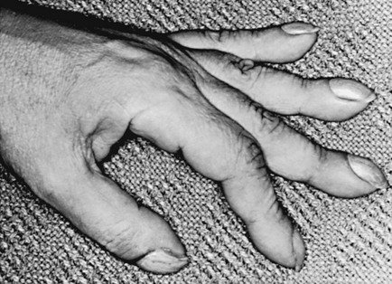
Clubbing is a change in the normal configuration of the nails and the distal phalanx of the fingers or toes (Fig. 3-1). Several features may be seen: (1) loss of the normal angle between the nail and the skin, (2) increased curvature of the nail, (3) increased sponginess of the tissue below the proximal part of the nail, and (4) flaring or widening of the terminal phalanx. Although several nonpulmonary disorders can result in clubbing (e.g., congenital heart disease with right-to-left shunting, endocarditis, chronic liver disease, inflammatory bowel disease), the most common causes clearly are pulmonary. Occasionally, clubbing is familial and of no clinical significance. Carcinoma of the lung (or mesothelioma of the pleura) is the single leading etiologic factor. Other pulmonary causes include chronic intrathoracic infection with suppuration (e.g., bronchiectasis, lung abscess, empyema) and some types of interstitial lung disease. Uncomplicated chronic obstructive lung disease is not associated with clubbing, so the presence of clubbing in this setting should suggest coexisting malignancy or suppurative disease.


Clubbing may be accompanied by hypertrophic osteoarthropathy, characterized by periosteal new bone formation, particularly in the long bones, and arthralgias and arthritis of any of several joints. With coexistent hypertrophic osteoarthropathy, either pulmonary or pleural tumor is the likely cause of the clubbing, because hypertrophic osteoarthropathy is relatively rare with the other causes of clubbing.


The mechanism of clubbing and hypertrophic osteoarthropathy is not clear. It has been observed that clubbing is associated with an increase in digital blood flow, whereas the osteoarthropathy is characterized by an overgrowth of highly vascular connective tissue. Why these changes occur is a mystery. One interesting theory suggests an important role for stimuli coming through the vagus nerve, because vagotomy frequently ameliorates some of the bone and nail changes. Another theory proposes that megakaryocytes and platelet clumps, bypassing the pulmonary vascular bed and affecting the peripheral systemic circulation, release growth factors responsible for the soft-tissue changes of clubbing.


Cyanosis, the second extrapulmonary physical finding arising from lung disease, is a bluish discoloration of the skin (particularly under the nails) and mucous membranes. Whereas oxygenated hemoglobin gives lighter skin and all mucous membranes their usual pink color, a sufficient amount of deoxygenated hemoglobin produces cyanosis. Cyanosis may be either generalized, owing to a low PO2 or low systemic blood flow resulting in increased extraction of oxygen from the blood, or localized, owing to low blood flow and increased O2 extraction within the localized area. In lung disease, the common factor causing cyanosis is a low PO2, and several different types of lung disease may be responsible. The total amount of hemoglobin affects the likelihood of detecting cyanosis. In the anemic patient, if the total quantity of deoxygenated hemoglobin is less than the amount needed to produce the bluish discoloration, even a very low PO2 may not be associated with cyanosis. In the patient with polycythemia, in contrast, much less depression of PO2 is necessary before sufficient deoxygenated hemoglobin exists to produce cyanosis.


Chest Radiography


The chest radiograph, which is largely taken for granted in the practice of medicine, is used not only in evaluating patients with suspected respiratory disease but also sometimes in the routine evaluation of asymptomatic patients. Of all the viscera, the lungs are the best suited for radiographic examination. The reason is straightforward: air in the lungs provides an excellent background against which abnormalities can stand out. Additionally, the presence of two lungs allows each to serve as a control for the other so that unilateral abnormalities can be more easily recognized.


A detailed description of interpretation of the chest radiograph is beyond the scope of this text. However, a few principles can aid the reader in viewing films presented in this and subsequent chapters.


First, the appearance of any structure on a radiograph depends on the structure’s density; the denser the structure, the whiter it appears on the film. At one extreme is air, which is radiolucent and appears black on the film. At the other extreme are metallic densities, which appear white. In between is a spectrum of increasing density from fat to water to bone. On a chest radiograph, the viscera and muscles fall within the realm of water density tissues and cannot be distinguished in radiographic density from water or blood.


Second, in order for a line or an interface to appear between two adjacent structures on a radiograph, the two structures must differ in density. For example, within the cardiac shadow, the heart muscle cannot be distinguished from the blood coursing within the chambers because both are of water density. In contrast, the borders of the heart are visible against the lungs because the water density of the heart contrasts with the density of the lungs, which is closer to that of air. However, if the lung adjacent to a normally denser structure (e.g., heart or diaphragm) is airless, either because of collapse or consolidation, the neighboring structures are now both of the same density, and no visible interface or boundary separates them. This principle is the basis of the useful silhouette sign. If an expected border with an area of lung is not visualized or is not distinct, the adjacent lung is abnormal and lacks full aeration.


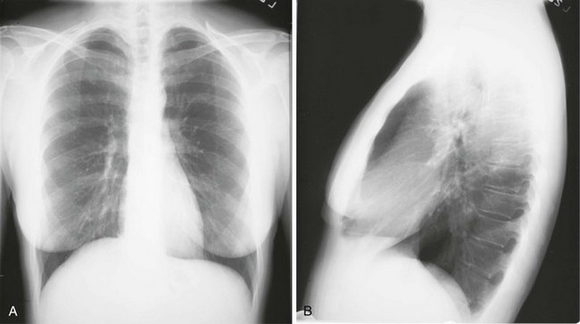
Chest radiographs usually are taken in two standard views—posteroanterior (PA) and lateral (Fig. 3-2). For a PA film, the x-ray beam goes from the back to the front of the patient, and the patient’s anterior chest is adjacent to the film. The lateral view is taken with the patient’s side against the film, and the beam is directed through the patient to the film. If a film cannot be taken with the patient standing and the chest adjacent to the film, as in the case of a bedridden patient, then an anteroposterior view is taken. For this view, which is generally obtained using a portable chest radiograph machine in the patient’s hospital room, the film is placed behind the patient (generally between the patient’s back and the bed), and the beam is directed through the patient from front to back. Lateral decubitus views, either right or left, are obtained with the patient in a side-lying position, with the beam directed horizontally. Decubitus views are particularly useful for detecting free-flowing fluid within the pleural space and therefore are often used when a pleural effusion is suspected.


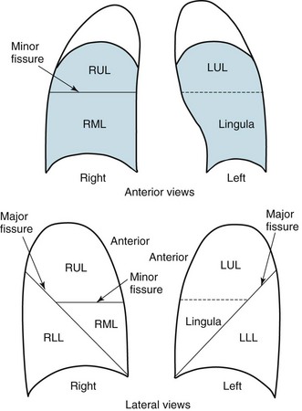
Knowledge of radiographic anatomy is fundamental for interpretation of consolidation or collapse (atelectasis) and for localization of other abnormalities on the chest film. Lobar anatomy and the locations of fissures separating the lobes are shown in Figure 3-3. Localization of an abnormality often requires information from both the PA and lateral views, both of which should be taken and interpreted when an abnormality is being evaluated. As can be seen in Figure 3-3, the major fissure separating the upper (and middle) lobes from the lower lobe runs obliquely through the chest. Thus it is easy to be misled about location on the basis of the PA film alone; a lower lobe lesion may appear in the upper part of the chest, whereas an upper lobe lesion may appear much lower in position.


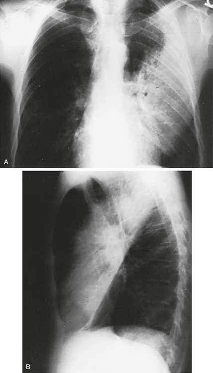
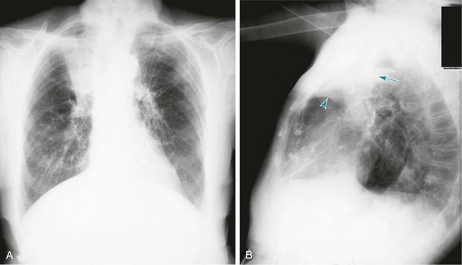
When a lobe becomes filled with fluid or inflammatory exudate, as in pneumonia, it contains water rather than air density and therefore is easily delineated on the chest radiograph. With pure consolidation the lobe does not lose volume, so it occupies its usual position and retains its usual size. An example of lobar consolidation on PA and lateral radiographs is shown in Figure 3-4.


In contrast, when a lobe has airless alveoli and collapses, it not only becomes more dense but also has features of volume loss characteristic for each individual lobe. Such features of volume loss include change in position of a fissure or the indirect signs of displacement of the hilum, diaphragm, trachea, or mediastinum in the direction of the volume loss (Fig. 3-5). A common mechanism of atelectasis is occlusion of the airway leading to the collapsed region of lung, caused, for example, by a tumor, aspirated foreign body, or mucous plug. All the aforementioned examples reflect either pure consolidation or pure collapse. In practice, however, a combination of these processes often occurs, leading to consolidation accompanied by partial volume loss.


When the chest film shows a diffuse or widespread pattern of increased density within the lung parenchyma, it often is useful to characterize the process further, depending on the pattern of the radiographic findings. The two primary patterns are interstitial and alveolar. Although the naming of these patterns suggests a correlation with the type of pathologic involvement (i.e., interstitial, affecting the alveolar walls and the interstitial tissue; alveolar, involving filling of the alveolar spaces), such radiographic-pathologic correlations are often lacking. Nevertheless, many diffuse lung diseases are characterized by one of these radiographic patterns, and the particular pattern may provide clues about the underlying type or cause of disease.


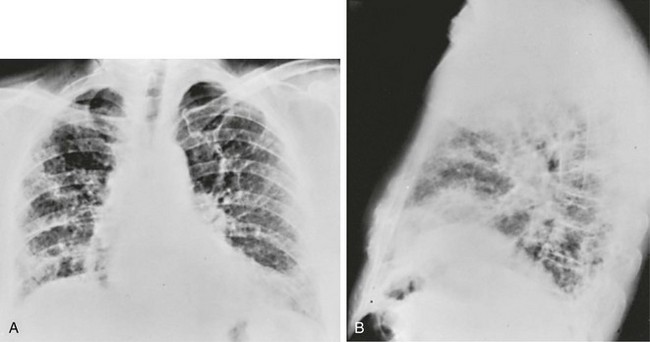
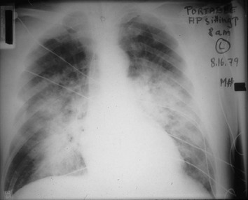
An interstitial pattern generally is described as reticular or reticulonodular, consisting of an interlacing network of linear and small nodular densities. In contrast, an alveolar pattern appears fluffier, and the outlines of air-filled bronchi coursing through the alveolar densities are often seen. This latter finding is called an air bronchogram and is due to air in the bronchi being surrounded and outlined by alveoli that are filled with fluid. This finding does not occur with a purely interstitial pattern. Examples of chest radiographs that show diffuse abnormality as a result of interstitial disease and alveolar filling are shown in Figures 3-6 and 3-7, respectively.


Two additional terms used to describe patterns of increased density are worth mentioning. A nodular pattern refers to the presence of multiple discrete, typically spherical, nodules. A uniform pattern of relatively small nodules several millimeters or less in diameter is often called a miliary pattern, as can be seen with hematogenous (bloodborne) dissemination of tuberculosis throughout the lungs. Alternatively, the nodules can be larger (e.g., > 1 cm in diameter), as seen with hematogenous metastasis of carcinoma to the lungs. Another common term is ground-glass, used to describe a hazy, translucent appearance to the region of increased density. Although the term can be used to describe a region or a pattern of increased density on a plain chest radiograph, it is more commonly used when describing abnormalities seen on computed tomography (CT) of the chest.


Although the preceding focus on some typical abnormalities provides an introduction to pattern recognition on a chest radiograph, the careful examiner must also use a systematic approach in analyzing the film. A chest radiograph shows not only the lungs; radiographic examination also may reveal changes in bones, soft tissues, the heart, other mediastinal structures, and the pleural space.


Computed Tomography


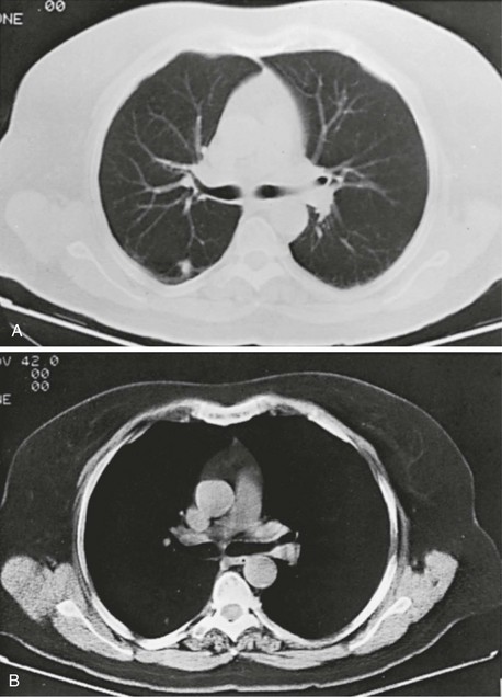
Compared with the plain chest radiograph, CT of the chest provides greater anatomic detail but is more expensive and exposes patients to a significantly higher dose of radiation. With this technique, a narrow beam of x-rays is passed through the patient and sensed by a rotating detector on the other side of the patient. The beam is partially absorbed within the patient, depending on the density of the intervening tissues. Computerized analysis of the information received by the detector allows a series of cross-sectional images to be constructed (Fig. 3-8). Use of different “windows” allows different displays of the collected data, depending on the densities of the structures of interest. With the technique of helical (spiral) CT scanning, the entire chest is scanned continuously (typically during a single breathhold and using multiple detectors) as the patient’s body is moved through the CT apparatus (the gantry).


CT is particularly useful for detecting subtle differences in tissue density that cannot be distinguished by conventional radiography. In addition, the resolution of the images and the cross-sectional views obtained from the slices provide better definition and more precise location of abnormalities.


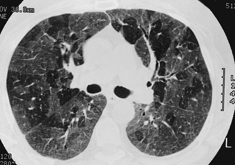
Chest CT is used extensively in evaluating pulmonary nodules and the mediastinum. It is also quite valuable in characterizing chest wall and pleural disease. As the technology has advanced, CT has become progressively more useful in the diagnostic evaluation of various diseases affecting the pulmonary parenchyma and the airways. With high-resolution CT, the thickness of individual cross-sectional images is reduced to 1 to 2 mm instead of the traditional 5 to 10 mm. As a result, exceptionally fine detail can be seen, allowing earlier recognition of subtle disease and better characterization of specific disease patterns (Fig. 3-9).


In the last decade, computed tomographic angiography (CTA) has become important in the diagnosis of pulmonary emboli. This technique, in which the pulmonary arterial system is visualized by helical CT scanning following injection of radiographic contrast into a peripheral vein, has been increasingly used in place of both perfusion lung scanning and traditional pulmonary angiography (see later.) Its use is attractive because CTA is more likely to be diagnostic than perfusion scanning, and it is less invasive than traditional pulmonary angiography. Although CTA may not be as sensitive as traditional angiography for detecting emboli in relatively small pulmonary arteries, ongoing improvements in CT scanner technology have led to better identification of clots in progressively smaller pulmonary arteries.


Sophisticated software protocols now allow images obtained by CT scanning to be reconstructed and presented in any plane that best displays the abnormalities of interest. Additionally, three-dimensional images are produced from the data acquired by CT scanning. For example, a three-dimensional view of the airways can be displayed in a manner resembling what is seen inside the airway lumen during bronchoscopy (described later in this chapter). This methodology creates an imaging tool that has been dubbed virtual bronchoscopy.


Magnetic Resonance Imaging


Another technique available for evaluation of intrathoracic disease is magnetic resonance imaging (MRI). The physical principles of MRI are complicated and beyond the training of most physicians and students, but are discussed here briefly. The interested reader is referred to other sources for an in-depth discussion of MRI (see References). In brief, the technique depends on the way nuclei within a stationary magnetic field change their orientation and release energy delivered to them by a radiofrequency pulse. The time required to return to the baseline energy state can be analyzed by a complex computer algorithm, and a visual image created.


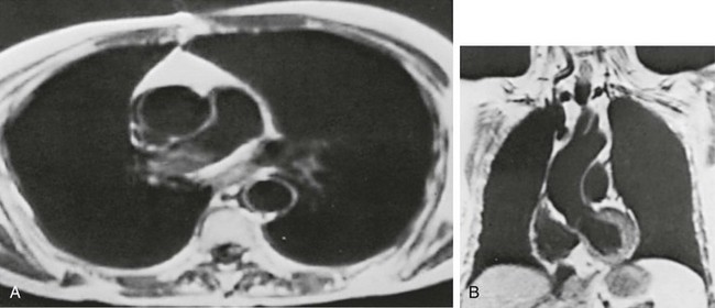
MRI has several important features in the evaluation of intrathoracic disease. First, flowing blood produces a “signal void” and appears black, so blood vessels can be readily distinguished from nonvascular structures without the need to use intravenous contrast agents. Second, images can be constructed in any plane so that the information obtained can be displayed as sagittal, coronal, or transverse (cross-sectional) views. Third, differences can be seen between normal and diseased tissues that are adjacent to each other, even when they are of the same density and therefore cannot be distinguished by routine radiography or CT. Some of these features are illustrated in Figure 3-10.


Stay updated, free articles. Join our Telegram channel

Jun 12, 2016 | Posted by in RESPIRATORY | Comments Off on Evaluation of the Patient with Pulmonary Disease

Full access? Get Clinical Tree

Get Clinical Tree app for offline access