10 Evaluation of Coronary Blood Flow by Echo Doppler
Background
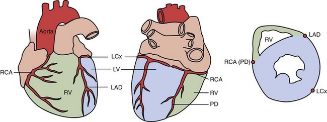
Anatomy (Fig. 10-1)
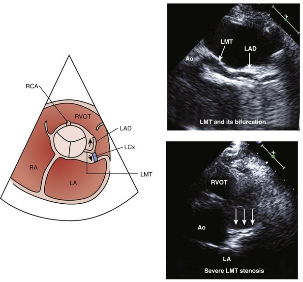
• The left main coronary artery originates from the left coronary sinus of the aortic root, and then bifurcates into the left anterior descending (LAD) and the left circumflex (LCx) arteries.
• The LAD runs over the anterior interventricular sulcus and extends apically, ending distal to the apex.
• The LCx is covered by the left atrial appendage in its proximal portion, and then takes position in the left atrioventricular sulcus.
• The right coronary artery (RCA) originates from the right coronary sinus of the aortic root, and goes into the right atrioventricular sulcus. The posterior descending artery (PDA) runs into the posterior interventricular sulcus.
Pathophysiology
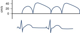
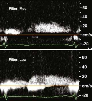
• Normal coronary flow is characterized by a biphasic flow profile, consisting of small systolic and large diastolic flow (Fig. 10-2).
• Doppler spectral tracings of coronary flow provide some useful clinical information such as coronary flow direction, flow velocity, and flow pattern.
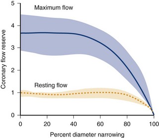
• Coronary flow reserve (CFR) is expressed as a ratio of maximum flow to resting flow, with the normal response being an increase in flow to four times baseline (Fig. 10-3).
• CFR decreases to only twice baseline at approximately 75% diameter stenosis, which indicates significant myocardial stenosis.
Anatomic Imaging and Physiologic Data Acquisition
Detection of Coronary Flow Signal by Color Doppler
Left Main Trunk, Proximal Left Anterior Descending Artery, and Left Circumflex Artery (Use Low-Frequency Transducer)
• The left main trunk (LMT) and its bifurcation can be detected at the level of the left coronary sinus, in a short axis view (Fig. 10-7).
• The proximal RCA can be identified at the level of the right coronary sinus in a short axis or long axis view.
Middle to Distal Portion of the Left Anterior Descending Artery
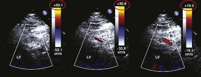
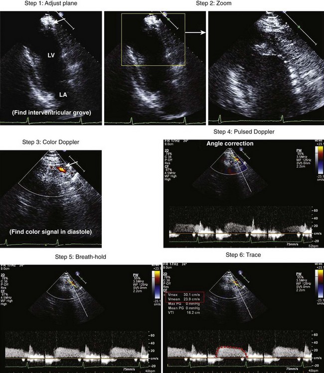
• The middle portion of the LAD can be identified in short axis images of the left ventricle (LV), located in the anterior interventricular sulcus (Fig. 10-8).
• Rotate the transducer counterclockwise to image the LV in a long axis view along the intraventricular sulcus under the guidance of color Doppler flow mapping.
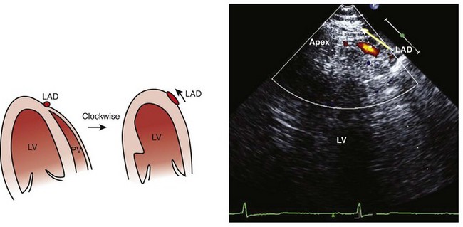
• The distal portion of the LAD can be found in the apical long axis view at the intraventricular sulcus, again as a circular cross section containing the color Doppler flow signal.
• Rotate the transducer clockwise (toward the two-chamber view position) to detect the flow signal in the long axis view.
• Coronary flow detection in the distal portion of the LAD is important because CFR should be measured distal to a stenosis.
Right Posterior Descending Artery
• Obtain an apical two-chamber image of the LV and angle the ultrasound beam superiorly to image the posterior interventricular sulcus using a low-frequency transducer (Fig. 10-9).
• Examine this area using color Doppler flow mapping to locate the right posterior descending coronary artery.
Stay updated, free articles. Join our Telegram channel

Full access? Get Clinical Tree


