, Fraser Munro1, Jimmy Lam1 and Gordon MacKinlay1
(1)
Department of Paediatric Surgery, Royal Hospital for Sick Children, Edinburgh, UK
Abstract
Thoracoscopic repair of esophageal atresia (OA) and tracheoesophageal fistula (TOF) is one of the most advanced and technically demanding minimally invasive surgery (MIS) operations in pediatric surgery. Since the first report by Rothenberg and Lobe in 1999 [1], there has been much investment in the development of this operation. There remains much debate about the benefits and risks of the thoracoscopic approach compared with those of open surgery. Nevertheless it is now performed by those with sufficiently advanced MIS skills with good outcomes.
Keywords
Oesophageal atresiaTracheoesophageal fistulaThoracoscopyNeonates10.1 General Information
Thoracoscopic repair of esophageal atresia (OA) and tracheoesophageal fistula (TOF) is one of the most advanced and technically demanding minimally invasive surgery (MIS) operations in pediatric surgery. Since the first report by Rothenberg and Lobe in 1999 [1], there has been much investment in the development of this operation. There remains much debate about the benefits and risks of the thoracoscopic approach compared with those of open surgery. Nevertheless it is now performed by those with sufficiently advanced MIS skills with good outcomes.
10.2 Working Instruments
3- (and 5-mm) Ports
3- or 5-mm 30-degree Telescope (short)
3-mm Needle holders (preferably left- and right-handed)
3-mm Hook diathermy
or Bipolar forceps (e.g., LigaSure)
3-mm Johan forceps (×2)
3-mm Maryland forceps
3-mm Metzenbaum and hook scissors
10.3 Positioning, Port Siting, and Ergonomic Considerations
The approach is through the right side of the chest with the patient in a semi-prone position, with the right side elevated 30–45°. This allows gravity to assist with lung retraction away from the posterior mediastinum. The right arm is positioned above the head with suitable padding.
The primary port position is just below the tip of the scapula, in the fourth or fifth intercostal space in the midaxillary line (Fig. 10.1). The working ports are placed under vision in the anterior and posterior axillary lines. Lung collapse is achieved by low pressure CO2 pneumothorax at 4–6 mmHg pressure. This takes a few minutes to achieve and can be aided by gentle compression of the lung with instruments.
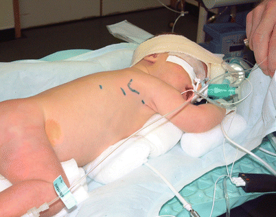
Fig. 10.1
Siting of ports. The scapula is outlined. The primary port site is shown at the interspace below the tip of the scapula
10.4 Relevant Anatomy (Figs. 10.2 and 10.3)

Fig. 10.2
Anatomy of the right posterior mediastinum as seen on entering. The azygos vein is seen crossing the upper mediastinum (over the upper aorta, which is just coming into view at this level) to enter the SVC. The vagus nerve is seen and is a good landmark to identify the lower pouch and eventually the fistula

Fig. 10.3
View of the uppermost part of the mediastinum showing the upper pouch (made more visible by the presence of the Replogle tube). Again note the right vagus nerve lying between the upper esophagus and trachea. The azygos vein is crossing over the esophageal gap
10.5 Surgical Technique (Figs. 10.4, 10.5, 10.6, 10.7, 10.8, 10.9, 10.10, 10.11, 10.12, 10.13, 10.14, 10.15, 10.16, 10.17, 10.18, 10.19, 10.20, 10.21 and 10.22)

Fig. 10.4
Identification of the lower pouch and fistula. After entering the chest, identification of the azygos vein and the vagus nerve helps to orientate the anatomy and identify the esophagus (lower pouch)

Fig. 10.5


Dissection of the azygos vein if needed. It may need to be divided if it obscures the approach to the lower pouch or fistula, but it is not absolutely necessary. The pleura is incised over the azygos vein to expose it’s surface and then freed by blunt dissection until a sufficient length is achieved for safe coagulation and division. Note the upper pouch seen during this dissection
Stay updated, free articles. Join our Telegram channel
Full access? Get Clinical Tree
