(1)
Department of Cardiovascular and Endovascular Surgery, The Arizona Heart Foundation, Phoenix, AZ, USA
Abstract
Aortic coarctation has been recognized since the late 1700s [1], but the first and largest post-mortem series appeared in 1928 [2]. Nevertheless, the condition was not commonly diagnosed clinically until the early 1930s. Although de novo lesions in adults are rare, primary aortic coarctation has been documented in patients over a wide range of ages and with varying degrees of severity—in general, the condition presents most commonly in infancy. Aortic coarctation is considered significant if an invasively determined pressure gradient is >20 mmHg at rest or >30 mmHg after exercise in adolescents and adults. Untreated coarctation can cause left ventricular pressure overload and left ventricular hypertrophy, premature coronary artery disease, and, eventually, heart failure.
Introduction
Aortic coarctation has been recognized since the late 1700s [1], but the first and largest post-mortem series appeared in 1928 [2]. Nevertheless, the condition was not commonly diagnosed clinically until the early 1930s. Although de novo lesions in adults are rare, primary aortic coarctation has been documented in patients over a wide range of ages and with varying degrees of severity—in general, the condition presents most commonly in infancy. Aortic coarctation is considered significant if an invasively determined pressure gradient is >20 mmHg at rest or >30 mmHg after exercise in adolescents and adults. Untreated coarctation can cause left ventricular pressure overload and left ventricular hypertrophy, premature coronary artery disease, and, eventually, heart failure.
Successful surgical correction of coarctation of the aorta by resection with end-to-end anastomosis was first described by Crafoord and Nylin in 1945 [3]. Until fairly recently, open surgical repair using either a left thoracotomy with end-to-end anastomosis or end-to-side anastomosis was considered “gold-standard” treatment of coarctation [4–7]. Despite good long-term success, recurrence of coarctation is considerable—as high as 15 % [4–8]. Percutaneous balloon angioplasty was introduced as a treatment in 1981 [9], and we performed one of the early stenting procedures at The Arizona Heart Institute in 1995 [10]. In recent years, stenting (sometimes combined with hybrid repair) has become an accepted treatment for aortic coarctation, reserving surgery for those patients not suited for the endovascular approach or in whom it has failed [11–15].
Patient Selection
Because de novo lesions are rare in adults, most patients who are candidates for endovascular repair have had a previous intervention. Factors to be considered when deciding the most appropriate form of management include age, aortic morphology, any previous surgical or endovascular treatment, and the interventionist’s experience. Arch hypoplasia may cause a residual gradient following coarctation repair, particularly if it is severe and this condition represents a significant challenge for successful repair.
Approximately 10 % of patients who underwent surgical repair of neonatal aortic coarctation will develop recurrence as an adult and present with associated aortic diseases. Open repair can be challenging because of complex aortic pathology with the recoarctation, intercostal collateral circulation, and pleural scar tissue from the original operative approach.
Early repair of native coarctation is associated with a lower risk of late hypertension and improved survival, but the risk of recoarctation is higher. Recoarctation with aneurysm or pseudoaneurysm formation is fairly common. Aneurysms may develop at the site of surgical repair or within the proximal aorta and often these are best treated with endovascular therapy; aneurysmal disease distal to a recoarctation is fairly common. Untreated aneurysms carry a considerable risk of aortic rupture. With late repair, there is a greater risk of recurrent hypertension and a lower overall risk of recoarctation; nevertheless, survival and hypertension may still be improved by treating patients who are first diagnosed as adults.
Indications
The presentation of coarctation is dependent on the severity of obstruction and associated cardiac lesions; in general, those diagnosed as infants exhibit congestive heart failure, severe acidosis, and ischemia of the lower extremities. Adolescent and adult patients often present with hypertension that is difficult to control, headaches, nosebleeds, leg cramps, muscle weakness, claudication, ischemia of the bowel and lower extremities, and neurologic changes.
Technique
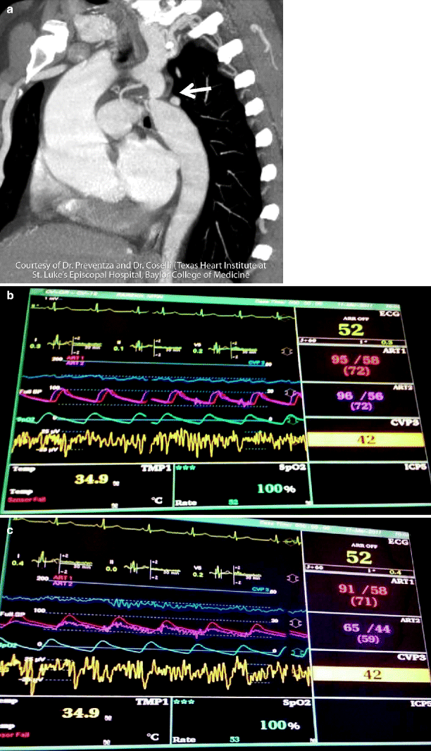
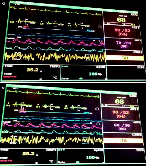
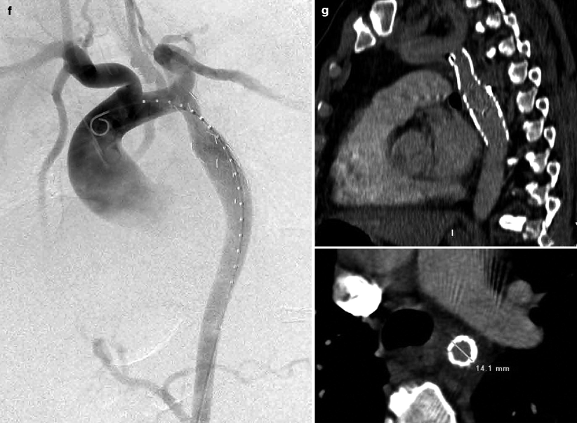
The goal of an endovascular approach to treat coarctation of the aorta is to restore anatomical relief of the obstruction resulting in reduction of the aortic gradient pressures. This concept is exemplified well in Fig. 42.1, which was contributed by one of my former fellows. Endovascular approaches are preferable to open surgery in the vast majority of patients with recoarctation, and balloon angioplasty and stent implantations are widely used [11, 16]; endoluminal grafting (including covered stents) may also be appropriate in selected cases [14]. Both covered and uncovered stents can be used to achieve satisfactory results. At our institution, we use endovascular techniques to provide relief from obstruction at the coarctation site and improve pressures proximally and distally as well as to exclude pericoarctation aneurysms or dissection to minimize the risk of rupture. The average duration of hospitalization following stenting of aortic coarctation is about 2 days, whereas open surgery may require a stay as long as 2 weeks. We strive to avoid disruption of blood flow to the left upper extremity and vertebral circulation, but when subclavian artery coverage is required, a carotid-subclavian bypass prevents vascular compromise to the left upper extremity. All patients with documented coarctation-associated pseudoaneurysms routinely undergo preoperative computed tomography (CT) angiography and Doppler studies to delineate the anatomical and physiological status of their carotid and vertebral arteries and its branches. If the operative procedure entails coverage of the left subclavian artery, we perform elective left carotid-subclavian bypass grafting whenever the right vertebral artery is abnormally small or diseased, or when there is evidence of an incomplete circle of Willis [12]. In the rare case in which the patient has had a left internal mammary artery bypass to the coronary circulation, the carotid-subclavian bypass is also necessary.


Fig. 42.1
(a) Angiogram showing narrow (arrow) recurrent coarctation distal to the left subclavian artery (Courtesy of Dr. Preventza and Dr. Coselli [Texas Heart Institute at St. Luke ’s Episcopal Hospital, Baylor College of Medicine]). (b) Pressure tracings using pigtail catheter proximal to coarctation. (c) Tracings distal to coarctation. (d) Tracings proximal to coarctation after stent and balloon angioplasty using 12 mm balloon. (e) Tracing distal to repair with no pressure gradient. (f) Comparison of pre- and postoperative angiograms following successful angioplasty. (g) Post-procedure CT examination showing good apposition of stent and satisfactory 14.1 mm residual aortic diameter
Angioplasty
Balloon angioplasty is a widely accepted treatment for patients with native and recurrent coarctation of the aorta in patients older than 6 months of age. The angioplasty procedure stretches and tears the intima and, in some cases, the media of the vessel wall. While ample luminal gain can be achieved, the correct balloon size is key in preventing elastic recoil and inordinate trauma to the vessel. In older patients with degenerative disease and calcification, great care must be taken when dilating the vessel. Indeed, safe use of angioplasty requires a delicate balance, as underdistension of the vessel may cause residual stenosis, and overdistension can result in aortic dissection, rupture, or aneurysm formation. In addition, neointimal proliferation and restenosis may result from vessel wall trauma following intervention. Because of these potential adverse events, stenting (rather than angioplasty alone) has become almost routine at our institution as well as others.
Stenting
Bare stents are generally used to ensure adequate aortic lumen reduction of the pressure gradient and a lower recurrence rate than that seen with angioplasty. Stenting addresses resulting vessel wall trauma from ballooning, prevents elastic recoil, and seals small dissections. Neointimal response and proliferation may be observed following stenting, but stent treatment of coarctation in older children, adolescents, and adults results in immediate hemodynamic benefit [10– 13, 15]; in larger vessels, the recurrence rate is very low. Our preference for stenting has been the Palmaz XL 10 series stent (Cordis, Miami Lakes, FL) since for a time it was the only large product available. The IntraStent Max (ev3 Endovascular Inc., Plymouth, MN), approved in 2002, offers another option; however, because of its rounded edges, it results in less pressure in the vessel wall, and migration may be a problem. Other stents, such as the Cheatham-Platinum stent (NuMED, Hopkinton, NY), appear to be highly effective in reducing the coarctation gradient and increasing lesion dilation in both native coarctation and recoarctation but are not yet available in the US [17].
< div class='tao-gold-member'>
Only gold members can continue reading. Log In or Register to continue
Stay updated, free articles. Join our Telegram channel

Full access? Get Clinical Tree


