Endocrine, Metabolic, and Electrolyte Responses to Cardiac Surgery and Cardiopulmonary Bypass


Endocrine, Metabolic, and Electrolyte Responses to Cardiac Surgery and Cardiopulmonary Bypass


Mark T. Nelson

Sarah K. Armour

John F. Butterworth IV


Surgical procedures performed with cardiopulmonary bypass (CPB) produce physiologic alterations not found in other major surgical procedures. During total CPB, the heart and lungs are not perfused and can neither secrete hormones nor make their normal contributions to drug metabolism. Exposure to the pump-oxygenator and its tubing traumatizes cellular blood elements, causes plasma proteins to be adsorbed and removed from the circulation, and stimulates an immune response, as is well described in other chapters in this volume. Hemodilution (from blood-free priming solutions) and anticoagulation alter blood concentrations of electrolytes, hormones, and serum proteins during CPB. Finally, moderate to profound hypothermia is often used, reducing the rates of biochemical reactions and further perturbing hormonal responses.

Certain features of extracorporeal perfusion contribute to the endocrine, metabolic, and electrolyte alterations. Nonpulsatile perfusion has been shown to change the distribution of flow both among and within organs. As a consequence, some hormonal alterations during CPB can be lessened or prevented by pulsatile perfusion. CPB increases “stress” hormones disproportionate to the apparent levels of physiologic disturbance, and it remains unclear which factor—hypothermia, hemodilution, decreased perfusion of endocrine glands, or denaturation of hormones by foreign surfaces—most contributes to these changes. Additionally, some hormone concentrations increase above normal levels after termination of bypass with the return of pulsatile normothermic perfusion to endocrine glands (1). Consistent with expectations, some data show that deeper planes of anesthesia attenuate or eliminate the exaggerated endocrine responses to CPB and may reduce mortality (2). Finally, spinal and epidural anesthesia and analgesia have been used during cardiac surgery, and these techniques inhibit the neuroendocrine response to cardiac surgery just as they do to abdominal and lower extremity surgery (3).

We find summarizing the literature regarding endocrine, metabolic, and electrolyte responses to CPB a difficult task. There are marked variations from center to center or study to study in patient populations, perfusion and cardioplegia techniques, perfusate temperatures, priming solutions, and anesthetic and adjuvant drugs. Earlier studies in which the hormone assays were not specific for intact, active hormones exacerbate the confusion. With these several concerns in mind, this chapter emphasizes the most recent studies in which contemporary anesthesia, cardioplegia, perfusion, and hormone measurement techniques were used.


PITUITARY HORMONES

The anterior portion of the pituitary gland secretes hormones that regulate the adrenal cortex, thyroid, ovaries, and testes. Several aspects of pituitary response (e.g., those related to the cortisol and thyroid axes) are considered in subsequent sections. Gonadotropin responses during CPB have not been reported using modern surgical or analytic techniques (4,5,6). However, Maggio et al. (5) found significant perioperative decreases in testosterone concentrations in men and increases in women undergoing cardiac surgery. Estradiol levels were conversely significantly increased in men and decreased in women.

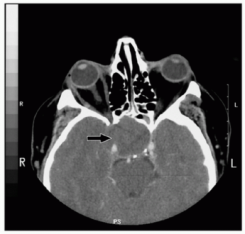
Pituitary apoplexy, a rare but potentially devastating complication, has been reported after CPB (7,8,9,10,11,12), typically in patients with pituitary adenomas. Rapid pituitary enlargement can compress parasellar structures such as the optic chiasm and ocular motor nerve resulting in varying combinations of ptosis, ophthalmoplegia, nonreactive and dilated pupils, decreased visual acuity, and visual field defects in addition to the characteristic hormonal deficits (13). Pituitary apoplexy presents only rarely as Addisonian crisis; more often oculomotor or visual disturbances or unexplained fever is the most common initial sign (8). Although ischemia, hemorrhage, and edema of the gland are usually assigned the blame for pituitary failure after bypass, no specific etiology has been identified in most patients. Anticoagulation alone has been associated with hemorrhage into pituitary adenomas, with both chronic oral and short-term heparin therapy (14); and there exists a gender bias with males outnumbering females in the ratio of 10 to 1 (15). The diagnosis can be confirmed with cranial computed tomography (CT) or magnetic resonance imaging (MRI) (Fig. 11.1). Hormonal replacement and prompt hypophysectomy are indicated, and experience suggests that the latter may be performed safely early after cardiac surgery (7,9,15). CPB alone does not lead to persisting hypopituitarism. When there is no
identifiable pituitary mass on CT or MRI, pituitary hormones are stable following CPB (16).
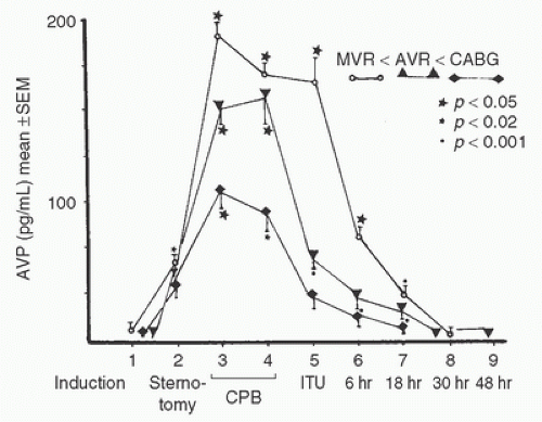


FIGURE 11.1. Cranial tomographic scan of a 56-year-old man 3 days after mitral valve repair. The patient presented with unilateral pupillary mydriasis, complete ophthalmoplegia, and loss of sensation in divisions I and II or cranial nerve V upon extubation several hours after his surgery. Note the mass in the sella turcica and bony erosion of the sphenoid “wing,” as indicated by the arrows. (From Meek EN, Butterworth J, Kon ND, et al. New onset of cranial nerve palsies immediately following mitral valve repair. Anesthesiology 1998;89:1580-1582, with permission.)


Vasopressin

Vasopressin, or antidiuretic hormone (ADH), secreted by the posterior pituitary gland, is a potent regulator of renal water excretion (17). At high concentrations, ADH may increase peripheral vascular resistance and decrease cardiac contractility and coronary blood flow (17,18). Animal studies have shown that infused arginine vasopressin (AVP) decreases cardiac oxytocin receptor expression and increases diastolic dysfunction in induced ischemia animal models (19,20). However, post-cardiac-surgery patients showed no cardiac dysfunction with AVP infusions (21,22). Similarly, in septic patients, those who received AVP required less vasopressors compared to controls while having no increase in mortality (23). ADH increases renal vascular resistance, reducing renal blood flow. ADH stimulates the release of the von Willebrand factor, perhaps improving hemostasis during and after cardiac surgery (see Chapter 22). Stimuli provoking ADH release include increased plasma osmolality, decreased blood volume or blood pressure, hypoglycemia, angiotensin, stress, and pain (17). General anesthesia and surgery are associated with moderate increases in ADH (24,25), and angiotensin-converting enzyme (ACE) inhibitors, which are commonly administered to cardiac surgical patients, have been associated with the syndrome of inappropriate antidiuretic hormone (SIADH) secretion (26). Cardiac surgery with CPB is associated with striking increases in ADH concentration, far above those seen during other major surgical procedures, and these effects may persist for hours postoperatively (25,27,28,29,30) (Fig. 11.2).


FIGURE 11.2. Plasma concentration of arginine vasopressin (AVP) during nonpulsatile bypass for mitral valve replacement (MVR, n = 8), aortic valve replacement (AVR, n = 5), or coronary artery bypass grafting (CABG, n = 5). Data are presented as means ± SEM. As indicated, measurements were obtained at (1) anesthesia induction, (2) sternotomy, (3) 10 minutes after initiation of cardiopulmonary bypass, (4) 10 minutes before termination of cardiopulmonary bypass, (5) upon arrival in the critical care unit, (6) 6 hours after bypass, (7) 18 hours after bypass, (8) 30 hours after bypass, and (9) 48 hours after bypass. All three groups of patients demonstrated significant increases in AVP concentrations during bypass. Only at sample 5 did the mitral valve patients demonstrate significantly greater AVP concentration than the CABG patients. p values on the figure indicate comparisons between sample 1 and subsequent samples in the same surgical group. (From Kaul TK, Swaminathan R, Chatrath RR, et al. Vasoactive pressure hormones during and after cardiopulmonary bypass. Int J Artif Organs 1990;13:293-299, with permission.)

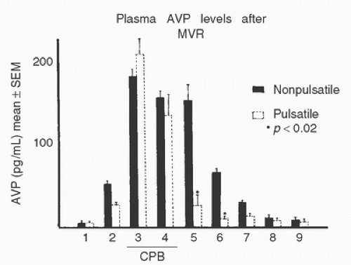
The exaggerated ADH response to CPB could be initiated by any number of stimuli, including the decrease in circulating blood volume upon initiating bypass. Left atrial pressure decreases markedly, especially with left-ventricular venting, thereby simulating volume depletion, which is a potent stimulus for ADH release. The transient hypotension normally occurring at the onset of bypass could stimulate increased ADH secretion. Pulsatile perfusion during CPB attenuates the exaggerated ADH response, particularly after bypass, but does not eliminate it (28,30,31) (Fig. 11.3). Pulsatile perfusion does not seem to significantly increase urinary output, despite reduced ADH concentrations (30).

Preoperatively, ADH levels were increased in patients with low left-ventricular ejection fraction (EF) or higher New York Heart Association (NYHA) heart failure class, and higher preoperative ADH concentrations served paradoxically as a predictor for postoperative vasoplegia. Plasma ADH concentrations did not increase nearly as much postoperatively in the vasoplegic group (32,33). This finding is consistent with the concept that vasoplegic
syndrome is related to inappropriately reduced ADH concentrations in those patients that had increased concentrations preoperatively, which may reduce the ability to increase ADH as part of the stress associated with cardiac surgery (32). A reduced EF and/or chronic treatment with ACE inhibitors were independently associated with vasoplegia and with this relative postoperative ADH deficiency (33). This finding is consistent with studies in sepsis associating inappropriately diminished ADH levels in the presence of chronic stimuli to ADH secretion (34).


FIGURE 11.3. Effect of pulsatile (n = 5) or nonpulsatile (n = 8) perfusion on arginine vasopressin (AVP) responses to mitral valve replacement. Significant differences between the two groups were observed after cardiopulmonary bypass (sample 5 and later). In this study, pulsatile bypass did not attenuate AVP responses during coronary bypass or aortic valve replacement. (From Kaul TK, Swaminathan R, Chatrath RR, et al. Vasoactive pressure hormones during and after cardiopulmonary bypass. Int J Artif Organs 1990;13:293-299, with permission.)


TABLE 11.1. Blood pressure and plasma catecholamine concentrations during extracorporeal perfusion in patients undergoing aortocoronary bypass grafting


Time sequence


Before anesthesia


After intubation


On bypass


Core temperature (32°C)


Core temperature (28°C)


Core temperature (24°C)


Core temperature, °C


37.0 ± 0 36.1


± 0.5a


34.7 ± 0.4a


31.5 ± 0.5a


27.8 ± 0.4a


24.1 ± 0.2a


MAP, mm Hg


86 ± 3


76 ± 1


70 ± 4a


73 ± 3a


60 ± 3a


60 ± 2a


NE, pg/mL


287 ± 40


360 ± 94


416 ± 83


662 ± 172a


540 ± 153


312 ± 86


EPI, pg/mL


50 ± 15


29 ± 8


138 ± 47


506 ± 191a


267 ± 136


130 ± 62


Core temperature, rectal temperature; EPI, plasma epinephrine concentration; MAP, mean arterial pressure; NE, plasma norepinephrine concentration. Catecholamine concentrations were not corrected for hemodilution.


a p < 0.05 compared with preinduction values.


Source: Reed HL, Chernow B, Lake CR, et al. Alterations in sympathetic nervous system activity with intraoperative hypothermia during coronary artery bypass surgery. Chest 1989;95:616-622, with permission.


Certain anesthetic techniques, for example, maintenance of anesthesia with large doses of synthetic opioids (fentanyl or sufentanil) or with regional anesthesia, attenuate the hormonal responses associated with surgical procedures. Indeed, Kuitunen et al. (35) found that patients anesthetized with 50 µg/kg fentanyl demonstrated significantly lower AVP concentrations after CPB than patients who received a lighter plane of general anesthesia using inhaled enflurane. However, even opioid anesthesia will not completely ablate ADH release at the onset of CPB (29). Unfortunately, multiple studies provide conflicting data as to whether higher peak ADH concentrations occur during and after CPB in patients undergoing coronary artery surgery or valve surgery (27,28,30) (Fig. 11.2). In summary, ADH concentrations increase markedly during CPB irrespective of the anesthesia or perfusion technique, and the level of increase may associate with the likelihood that a patient will develop vasoplegia.


ADRENAL HORMONES


Catecholamines

The catecholamines epinephrine and norepinephrine are products of the adrenal medulla and (in the latter case) of peripheral sympathetic and central nerve terminals. Marked elevations of plasma epinephrine and norepinephrine concentrations occurring during CPB underlie many hemodynamic sequelae of bypass, including peripheral vasoconstriction and shifts in intraorgan blood flow (31,36,37,38,39,40,41). With hypothermia, plasma epinephrine concentrations may increase as much as 10-fold over the prebypass concentrations; norepinephrine concentrations typically increase to a lesser extent (4-fold) (2,31,37,39), and deeper levels of hypothermia attenuate these (Table 11.1). In early studies, peak increases in both norepinephrine and
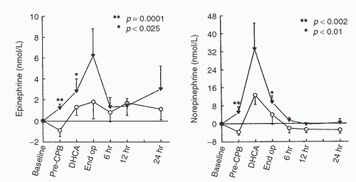
epinephrine occurred when the heart and lungs were excluded from the circulation (38,39,40). However, norepinephrine and epinephrine concentrations peaked at different times. In a later study, patients undergoing cardiac surgery were randomly assigned to have CPB with mild (34°C) or moderate (28°C) hypothermia. With both bypass temperatures, peak norepinephrine concentrations were observed after release of the aortic crossclamp and rewarming, whereas peak epinephrine concentrations were observed at the target hypothermic temperature (42). A more recent study demonstrated a biphasic plasma norepinephrine concentration response to nonpulsatile CPB, with concentrations peaking at aortic declamping and again 2 to 4 hours after surgery. Epinephrine concentrations did not show this pattern, nor was it observed during pulsatile CPB (40,41). Neonates, infants, and young children, much like adults, demonstrate marked increases in catecholamine concentrations during CPB (2,41,43,44,45) (Fig 11.4).

Deeper planes of general anesthesia (whether accomplished with larger doses of synthetic opioids, addition of a propofol infusion, higher concentrations of volatile anesthetic vapors, or addition of neuraxial anesthesia) significantly reduce the catecholamine concentrations of patients undergoing coronary artery bypass surgery compared with patients less deeply anesthetized (46,47,48). Furthermore, in critically ill neonates undergoing correction of congenital heart disease, deeper planes of general anesthesia from large intravenous doses of sufentanil not only produced lower catecholamine concentrations in response to CPB, but also reduced mortality compared with lighter planes of general anesthesia using halothane and morphine (2) (Fig 11.4). Consistent with these observations regarding anesthetic depth, infusion of propofol during CPB (4 mg/kg/hr) resulted in markedly reduced concentrations of epinephrine and norepinephrine compared with a single bolus injection of diazepam 0.1 mg/kg (47). Addition of thoracic epidural anesthesia to a “high-dose” fentanyl or sufentanil general anesthetic significantly reduced catecholamine concentrations during and after CPB relative to concentrations measured without thoracic epidural anesthesia (49,50) (Fig. 11.5). Similarly, patients undergoing CPB after “high spinal” intrathecal blockade had reduced levels of catecholamines compared to a control group, despite neither group receiving a high-dose opioid general anesthetic (51) (Fig. 11.5).


FIGURE 11.4. Perioperative changes in plasma epinephrine and norepinephrine in neonates undergoing cardiac surgery with either high-dose sufentanil (○; n = 30) or halothane-morphine (▼; n = 15) anesthesia. Pre-CPB, before bypass; DHCA, after deep hypothermic circulatory arrest; End op, end of operation; 6 hr, 12 hr, 24 hr, 6, 12, or 24 hours after operation. p values determined with Mann-Whitney U test. (From Anand KJS, Hickey PR. Halothane-morphine compared with highdose sufentanil for anesthesia and postoperative analgesia in neonatal cardiac surgery. N Engl J Med 1992;326:1-9, with permission.)

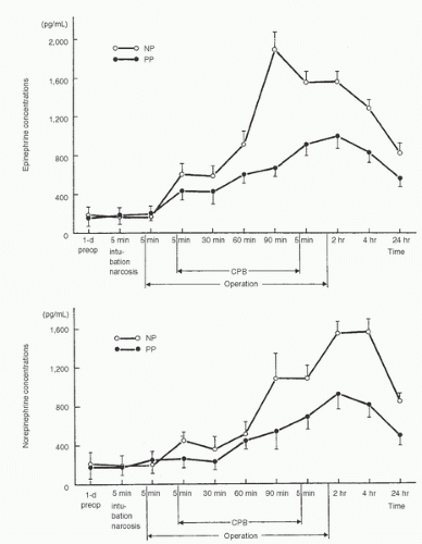
The effect of pulsatile perfusion on catecholamine concentrations during CPB remains controversial (31,52). Although early studies demonstrated that catecholamine concentrations were increased during CPB whether or not pulsatile perfusion was used (31), a more recent study of elective coronary surgery patients showed significant reductions in epinephrine and norepinephrine concentrations with pulsatile (vs. nonpulsatile) perfusion (40,52) (Fig. 11.6).

Some increase in catecholamine concentrations during and after CPB may be unavoidable with current anesthetic and surgical techniques; nevertheless, deeper planes of general anesthesia (either with larger doses of opioids or greater concentrations of inhaled general anesthetics) or addition of conduction anesthesia to general anesthesia can limit the increases.


Adrenal Cortical Hormones

Increased secretion of cortisol is one of the central features of the metabolic stress response. In the classic studies by Hume et al. (53) of patients undergoing major (noncardiac) surgery, cortisol concentrations rose quickly to a maximum and then slowly returned
to baseline 24 hours postoperatively. It is therefore not surprising that even “off-pump” coronary revascularization procedures are associated with increases in serum cortisol and other markers of the stress response despite the absence of the stress of CPB (54). It is apparent, however, that CPB modifies cortisol responses to surgery. Total plasma cortisol concentrations typically briefly decrease immediately upon initiation of bypass, likely as a consequence of hemodilution (55,56,57,58) (Fig. 11.7). During bypass, cortisol concentrations rise to values significantly above baseline (2,5,55,56,57,58,59,60). After CPB, patients exhibit markedly elevated concentrations of cortisol (both free and total) for more than 48 hours (58,59,60,61). In a recent study, cortisol increased 5-fold from baseline 2 hours after “on pump” coronary artery surgery versus a maximal 3-fold increase after “off pump” coronary artery surgery (60). Interestingly, the maximal increase occurred significantly earlier in the “off pump” group (4 hours after surgery) than in the “on pump” group (12 hours after surgery). The authors speculated that this finding resulted from differences in inflammatory processes between the two forms of coronary artery surgery. Cortisol levels slowly decreased thereafter in both groups.
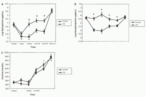


FIGURE 11.5. Effects of thoracic epidural anesthesia with bupivacaine 0.5% (▪, n = 8) versus control (♦, n = 9) on catecholamine concentrations measured during coronary artery surgery. All 17 patients studied received general anesthesia with sufentanil 20 µg/kg. Samples were obtained (1) before anesthesia, (2) after anesthesia induction, (3) after 30 minutes of surgery, (4) after 30 minutes of cardiopulmonary bypass (CPB), (5) after 60 minutes of CPB, (6) 1 hour after CPB, (7) 2 hours after CPB, (8) 4 hours after CPB, (9) 6 hours after CPB, and (10) 24 hours after CPB. *p < 0.05, **p < 0.01 for between-group differences. Adrenaline, epinephrine; NA, noradrenaline or norepinephrine. (From Lee TW, Grocott HP, Schwinn D, et al. High spinal anesthesia for cardiac surgery: effects on beta-adrenergic receptor function, stress response, and hemodynamics. Anesthesiology 2003;98:499-510, with permission.)

Leptin, an adipocyte-derived hormone, is thought to moderate the acute systemic inflammatory response to CPB and surgery and to interact importantly with the hypothalamic-pituitary-adrenal axis. Leptin binds to receptors in the hypothalamus and is known to affect energy metabolism. It has structural similarities to cytokines, it affects immunity, and it may modulate stress responses. Leptin concentrations decrease with cardiac surgery and CPB. “On pump” coronary surgical cases showed a more pronounced decrease in leptin compared to “off pump” coronary surgeries. Both groups also showed a subsequent increase in leptin 24 hours postoperatively. Leptin levels correlated inversely with plasma cortisol levels (60,62,63). Children undergoing surgical repair of congenital heart diseases with CPB demonstrated similar findings, with leptin concentrations decreasing during CPB and increasing afterward to peak at 12 hours postoperatively. Leptin concentrations were elevated in critically ill patients; thus elevations in leptin concentrations may serve as a marker for systemic inflammatory response syndrome (64).

Tinnikov et al. (65) studied 14 children undergoing repair of ventricular septal defects with deep hypothermia and circulatory arrest, but without CPB. Maximal perioperative concentrations of cortisol and minimal perioperative concentrations of cortisol binding globulin were recorded at the first assessment after circulatory arrest. Thus, hypothermia and circulatory arrest initiate a cortisol-stress response even in the absence of extracorporeal perfusion.

Cortisol responses during bypass appear to be temperature-dependent. Taggart et al. (66) showed that the increase in cortisol
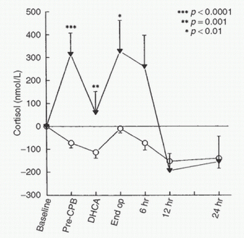
concentration during CPB can be blunted by perfusion with blood at 20°C compared to 28°C. Peak CPB cortisol concentrations were decreased by deeper planes of anesthesia in both adults and children (2,58,59) (Fig. 11.8). Winterhalter et al. (48) showed that when continuous remifentanil (0.25 µg/kg/min) was compared to intermittent bolus dose fentanyl (2.6 ± 0.3 mg/kg total dose) corticotropin (adrenocorticotropic hormone, ACTH), cortisol, and vasopressin were all significantly decreased. This result could be secondary to the specific agent (remifentanil vs. fentanyl), variations in the depth of anesthesia, or the steady state produced by continuous anesthetic infusions. Stenseth et al. (49) found that, compared with high-dose fentanyl anesthesia alone, high-dose fentanyl anesthesia plus thoracic epidural anesthesia delayed the increase in cortisol concentrations during coronary artery surgery and reduced concentrations during bypass. Similarly, Moore et al. (50) found that thoracic epidural anesthesia combined with sufentanil 20 µg/kg was associated with markedly lower cortisol concentrations as compared to sufentanil anesthesia alone. On the other hand, spinal anesthesia failed to attenuate cortisol responses (compared to intravenous general anesthesia) in children undergoing correction of congenital heart defects (3).


FIGURE 11.6. Effects of pulsatile (PP) and nonpulsatile (NP) perfusion on catecholamine responses in 30 patients undergoing coronary artery bypass grafting. Pulsatile perfusion significantly reduced both epinephrine and norepinephrine concentrations during bypass. Values are means ± SE. (From Minami K, Körner MM, Vyska K, et al. Effects of pulsatile perfusion on plasma catecholamine levels and hemodynamics during and after cardiac operations with cardiopulmonary bypass. J Thorac Cardiovasc Surg 1990;99:82-91, with permission.)

CPB modifies ACTH responses in surgical patients. In the previously mentioned study by Hume et al. (53), non-CPB surgical patients showed no increase in cortisol concentrations after an injection of ACTH, indicating that adrenal secretion of cortisol was
already maximal. Amado and Diago (67) observed a blunted response to corticotropin-releasing hormone during bypass, similar to responses seen in patients with hypothalamic corticotropin-releasing hormone deficiency. In contrast, an earlier study revealed that when patients undergoing extracorporeal perfusion received ACTH, cortisol concentrations increased (55).


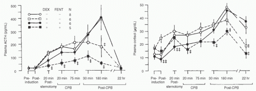
FIGURE 11.7. The effects of either enflurane or fentanyl anesthesia with or without dexamethasone treatment on cortisol and adrenocorticotropic hormone responses to cardiac surgery. All groups demonstrated significant increases in both cortisol and adrenocorticotropic hormone in response to surgery. The combination of fentanyl and dexamethasone significantly attenuated the adrenocorticotropic hormone response to surgery relative to the other three groups (p < 0.05 compared with the no dexamethasone, no fentanyl group; ‡‡p < 0.05 compared with the dexamethasone-treated, no fentanyl group). (From Raff H, Norton AJ, Flemma RJ, et al. Inhibition of the adrenocorticotropin response to surgery in humans: interaction between dexamethasone and fentanyl. J Clin Endocrinol Metab 1987;65:295-298, with permission.)


FIGURE 11.8. Cortisol responses during and after correction of congenital heart lesions with either halothane-morphine (n = 15, ▼) or sufentanil (n = 30, ○) anesthesia. The sufentanil-based technique significantly attenuated the “stress” response to cardiac surgery. (From Anand KJS, Hickey PR. Halothane-morphine compared with high-dose sufentanil for anesthesia and postoperative analgesia in neonatal cardiac surgery. N Engl J Med 1992;326:1-9, with permission.)

Taylor et al. (68) measured a progressive fall in ACTH concentrations during bypass, with a subsequent increase 1 hour after pulsatile perfusion was restored. Raff et al. (57) showed that, although neither high-dose fentanyl anesthesia nor dexamethasone 40 mg alone blunted the increase in ACTH concentration in response to CPB, concurrent administration of both agents significantly reduced the ACTH concentration (Fig. 11.7). In a 2011 study, Debono and colleagues (69) demonstrated that at least 25% of patients undergoing coronary artery bypass grafting (CABG) who had normal cosyntropin stimulation tests prior to surgery developed increased ACTH concentrations and decreased responses to cosyntropin (an ACTH derivative used in diagnostic testing) postoperatively. The clinical importance of this relative cortisol deficiency is unclear as postoperative outcomes were comparable regardless of ACTH concentrations or cosyntropin responses.

Unlike some other hormones, cortisol and ACTH responses to CPB generally have not been influenced by pulsatile perfusion. To be sure, one study found that total plasma cortisol rose during pulsatile bypass but fell dramatically in patients undergoing nonpulsatile perfusion (56). In another study, patients with and without pulsatile perfusion showed initial increases in cortisol, ACTH, and aldosterone, followed by a gradual decline in concentrations of all three hormones during bypass and then a subsequent increase in all three hormones after bypass perfusion (70). After correction for the effect of hemodilution, there was no decrease in calculated free cortisol concentrations and a slight increase in adrenocorticotropic hormone concentrations, irrespective of pulsatile versus nonpulsatile perfusion. In children with either pulsatile or nonpulsatile perfusion, Pollock et al. (71) found large increases in cortisol and ACTH during CPB, followed by a slow decline toward baseline concentrations of both hormones over 24 hours with both techniques.

Although there is no unequivocal evidence for adrenocortical hypofunction during or after CPB, the inflammatory response initiated by the triad of blood contact with the foreign surfaces of the extracorporeal membrane, reperfusion injury, and endotoxemia may be attenuated by large doses of exogenous glucocorticoids (72). This inflammatory response triggers tissue injury in the heart, kidneys, hemostatic system, and especially the lung, which
is the only organ exposed to the entire cardiac output (except during CPB). Early investigations have studied small numbers of cardiac surgery patients randomized to variable doses of different corticosteroids (most commonly 1 mg/kg dexamethasone or 30 mg/kg methylprednisolone) initiated at varying intervals between induction of anesthesia and the start of CPB (72). Overall, study results generally demonstrate an amelioration of the inflammatory response, with decreases in cytokine formation (tumor necrosis factor and the interleukin [IL]-1, -6, and -8) but inconsistent effects on C3a and elastase concentrations. Leukotrienes such as LTB4 are decreased in a dose-dependent fashion (72,73). IL-10, a cytokine with actions that are principally anti-inflammatory, demonstrates increased concentrations with steroid administration, supporting an anti-inflammatory effect (73). In addition, large doses of methylprednisolone can block upregulation of neutrophil integrin adhesion receptors, whereas dexamethasone decreases endothelial production of certain adhesion molecules (74). Clinically, glucocorticoid therapy may increase cardiac index and decrease systemic vascular resistance (75). Dietzman et al. (76) showed improvement in tissue perfusion and a decrease in peripheral vascular resistance when a large glucocorticoid dose was given just before CPB. Routine glucocorticoid supplementation has also been advocated as part of an accelerated recovery program (77), albeit without much supporting evidence. A small study has shown that cardiac surgery patients receiving glucocorticoids had shorter lengths of stay and improved quality of life as compared to patients not receiving glucocorticoids (78). The same research group found that steroid treatment reduced the duration of catecholamine support, length of intensive care unit (ICU) stay, and likelihood of postoperative atrial fibrillation (79). A 2013 meta-analysis that included 48 randomized controlled trials (RCTs) of patients receiving corticosteroids undergoing CPB failed to show any difference in the incidence of myocardial infarction, stroke, renal insufficiency, or death. The only outcome effect identified was a modest and heterogeneous decreased ICU and hospital length of stay in the groups receiving steroids (73).

In summary, current data nearly uniformly demonstrate large increases in cortisol and ACTH concentrations with initiation of CPB. These increases may be attenuated by deeper planes of general anesthesia or by addition of thoracic epidural (but apparently not spinal) anesthesia to general anesthesia as well as by continuous infusions of narcotic agents. Pulsatile perfusion does not appear to reduce these exaggerated responses. Moreover, it is not clear whether elevated corticosteroid concentrations during bypass are deleterious or beneficial.


GLUCOSE HOMEOSTASIS

Carbohydrate metabolism is regulated by insulin, glucagon, cortisol, growth hormone, and epinephrine, the concentrations of all of which are generally perturbed by surgery, CPB, and hypothermia. After the onset of CPB, blood glucose concentrations rise steadily if left untreated (80,81,82). Despite marked hyperglycemia, insulin concentrations decline from their control values during hypothermic bypass. Hyperglycemia, hypoinsulinemia, and insulin resistance are produced by hypothermic nonpulsatile CPB in adults (80,81,82). This catabolic response is greater during and following CABG with CPB than during and following off-pump coronary artery bypass, supporting the importance of CPB in this process (83). Strict normoglycemia can be maintained only with difficulty during hypothermic, nonpulsatile CPB in nondiabetic adults, even with large doses of insulin.

Studies performed in postoperative surgical patients have had a disproportionate influence in the intraoperative care of cardiac surgical patients. A prospective single center study in 2001 by van den Berghe et al. (84) in surgical ICU patients compared groups randomized to receive either insulin infusions to maintain “tight” glucose control (plasma concentrations 80-110 mg/dL) or insulin infusions maintaining plasma glucose <215 mg/dL demonstrated a substantial survival benefit in the “tight” glucose control group. Subsequent studies failed to demonstrate a survival benefit when plasma glucose concentrations were maintained at 80 to 110 mg/dL and in fact demonstrated an increased incidence of clinically significant hypoglycemia (85,86,87). A 2009 multicenter prospective randomized international trial (NICE-SUGAR) of medical and surgical ICU patients randomized to either a “tight” glucose control (plasma glucose concentrations 80-108 mg/dL) strategy or conventional control (glucose maintained <180 mg/dL) demonstrated decreased survival and increased incidence of hypoglycemia in the more restrictive (80-110 mg/dL plasma glucose concentration) group (88). This effect was present in both surgical and medical ICU patients. A 2010 meta-analysis by Mark and Prelser (89) of seven RCTs with over 11,400 patients comparing “tight” glucose control (glucose concentrations of 80-110 mg/dL) to less restrictive protocols failed to identify a survival benefit at 28 days, a decreased likelihood of renal replacement therapy, or a decreased incidence of blood-borne infections in the more restrictive (plasma glucose 80-110 mg/dL) group. The incidence of hypoglycemia was significantly increased in the more restrictive “tight” blood glucose group (89). In 2009, the Society of Thoracic Surgeons and in 2011 the American College of Physicians released guidelines supporting the use of less restrictive (glucose concentrations <180-200 mg/dL) blood glucose management strategies (90,91). In summary, current evidence suggests that attempting to maintain “strict” control of blood glucose (e.g., concentrations 80-120 mg/dL) does not improve outcome relative to a less restrictive strategy of maintaining blood glucose <180 mg/dL.

Counter-regulatory hormones decline from prebypass concentrations during hypothermic bypass (92). With rewarming in patients without diabetes, insulin concentrations rise spontaneously to appropriate high levels; nonetheless blood glucose concentration remains elevated. Normoglycemia is better preserved in children undergoing hypothermic CPB when washed red blood cells rather than conventional packed red blood cells (suspended in adenine-glucose-mannitol-saline) are used in the CPB priming solution (92,93). Many clinicians are likely unaware that blood glucose concentrations in packed red blood cells range from 400 to 700 mg/dL (93).


TABLE 11.2. Effect of nonpulsatile (n = 18) or pulsatile (n = 20) cardiopulmonary bypass on glucose metabolism


Sampling times


Postoperative (hr)


Variable


Group


Preoperative


CPB


1


5


24


48


Blood glucose, mg/dL


P


NP


83 ± 18


87 ± 27


206 ± 44a


254 ± 92a


253 ± 51a


281 ± 67a


235 ± 52a


271 ± 60a


125 ± 46


208 ± 72a


115 ± 27


149 ± 25


Immunoreactive insulin, mIU/L


P


NP


16 ± 9


13 ± 5


72 ± 39a


24 ± 15


71 ± 34a


29 ± 20b


89 ± 36a


40 ± 27b


72 ± 42


31 ± 23


64 ± 35


27 ± 18


Immunoreactive glucagon, ng/L


P


NP


98 ± 34


107 ± 45


150 ± 81


200 ± 96


148 ± 73


197 ± 88


202 ± 101


221 ± 79


225 ± 88


175 ± 80


220 ± 91


156 ± 66


CPB, cardiopulmonary bypass; NP, nonpulsatile CPB; P, pulsatile CPB.


Values are means ± SEM.


a p < 0.05 versus preoperative value;

b p < 0.05 versus group P.


Source: Nagaoka H, Innami R, Watanabe M, et al. Preservation of pancreatic beta cell function with pulsatile cardiopulmonary bypass. Ann Thorac Surg 1989;48:798-802, with permission.


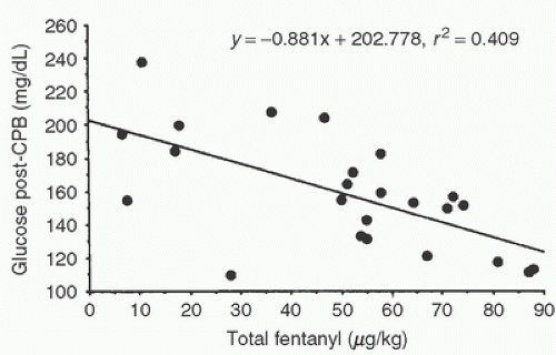
Concentrations of glucose, insulin, and glucagon are greater during hypothermic than normothermic CPB (82,94). Nagaoka et al. (80) compared pulsatile with nonpulsatile perfusion in patients undergoing cardiac surgery with moderate hypothermia (body temperature approximately 26°C). In both groups, blood glucose concentrations increased with CPB and rose further with hypothermia, reaching values greater than 200 mg/dL (Table 11.2). Blood glucose concentrations remained elevated for at least 5 hours postoperatively, but the patients receiving pulsatile perfusion showed a more rapid return to baseline glucose concentration than did patients receiving nonpulsatile perfusion. Insulin concentration, C peptide concentration, and the insulin-to-glucagon molar ratio increased significantly compared with baseline during pulsatile but not during nonpulsatile CPB. Type I (juvenile) diabetics require no greater doses of insulin to control blood glucose during CPB than do nondiabetic control subjects, whereas type II diabetics exhibit greater insulin resistance compared with type I diabetics and nondiabetic control patients during CPB (82). Children receiving deeper planes of general anesthesia demonstrated lower blood glucose concentrations upon termination of bypass than did children receiving lighter anesthetic techniques (2,95) (Fig. 11.9). Similarly, nondiabetic patients undergoing CABG with CPB that received epidural anesthesia required lower infusion doses of insulin and maintained lower blood glucose concentrations than did control patients that did not receive epidural anesthesia. Diabetic patients undergoing CABG with CPB who received epidural anesthesia had reduced blood glucose concentrations without a change in insulin requirements (96).

In adults undergoing coronary artery surgery and in children undergoing correction of congenital heart defects, growth hormone increased during and after CPB (97,98). The increase in growth hormone could be prevented using opioid general anesthetic techniques (98). The physiologic significance of this growth hormone response is unclear, because it can be inhibited by prior administration of somatostatin without an effect on glucose or glutamine metabolism. However, children whose catabolic hormonal responses waned by postoperative day 5 tended to have shorter lengths of stay (99).


FIGURE 11.9. Total fentanyl dose is inversely correlated with blood glucose concentrations in 24 children undergoing correction of congenital heart disease with hypothermic circulatory arrest (but without profound hypothermia or circulatory arrest). Blood samples were withdrawn within 30 minutes after cessation of bypass. p = 0.0007 for the slope of the regression line. (From Ellis DJ, Steward DJ. Fentanyl dosage is associated with reduced blood glucose in pediatric patients after hypothermic cardiopulmonary bypass. Anesthesiology 1990;72:812-815, with permission.)


NATRIURETIC PEPTIDES

Natriuretic peptides are a family of biologically active peptides first isolated from cardiac atria that include atrial natriuretic
peptide (ANP, A-type natriuretic peptide), brain natriuretic peptide (BNP, B-type natriuretic peptide), and C-type natriuretic peptide (CNP), among others (100). ANP and BNP are expressed in cardiac myocytes. The designation of BNP as “brain” natriuretic peptide derives from its initial discovery in porcine brain, but the role of BNP and ANP in the central nervous system remains unclear. ANP is produced and stored predominantly in the atrium. BNP is present in both atrial and ventricular tissue, but in myocardial disease states the ventricle becomes the primary source of BNP secretion. The nervous system and endothelial cells are the major sites of expression of CNP, which has significant paracrine vasodilatory effects. The heart is not a significant source of CNP. ANP is released in response to atrial distention, whereas BNP levels tend to be elevated in the presence of ventricular dysfunction. Other stimuli, some of which are commonly associated with CPB, may promote ANP and/or BNP release, including myocardial ischemia (101), catecholamines, endothelin-1, prostacyclin, and cytokines (102). Both peptides increase glomerular filtration, inhibit renin release, reduce aldosterone concentrations in blood, antagonize renal vasoconstrictors (such as vasopressin, norepinephrine, and angiotensin), and reduce arterial blood pressure. They regulate vascular volume by increasing sodium excretion and decreasing vasomotor tone (103). Within the heart natriuretic peptides regulate myocyte growth and inhibit fibroblast proliferation and extracellular matrix deposition. Natriuretic peptides have an anti-ischemic (preconditioning-like) function, and influence coronary endothelial and vascular smooth muscle proliferation and contractility through activation of guanylate cyclase (104,105).

Jun 7, 2016 | Posted by in RESPIRATORY | Comments Off on Endocrine, Metabolic, and Electrolyte Responses to Cardiac Surgery and Cardiopulmonary Bypass

Full access? Get Clinical Tree
Get Clinical Tree app for offline access