Electrophysiology
Cardiac cells
The heart is composed of thousands of cardiac cells. The cardiac cells are long and narrow, and divide at their ends into branches. These branches connect with branches of adjacent cells, forming a branching and anastomosing network of cells. At the junctions where the branches join together is a specialized cellular membrane of low electrical resistance, which permits rapid conduction of electrical impulses from one cell to another throughout the cell network. Stimulation of one cardiac cell initiates stimulation of adjacent cells and ultimately leads to cardiac muscle contraction.
There are two basic kinds of cardiac cells in the heart: the myocardial cells (or “working” cells) and the pacemaker cells. The myocardial cells are contained in the muscular layer of the walls of the atria and ventricles. The myocardial “working” cells are permeated by contractile filaments which, when electrically stimulated, produce myocardial muscle contraction. The primary function of the myocardial cells is cardiac muscle contraction, followed by relaxation. The pacemaker cells are found in the electrical conduction system of the heart and are primarily responsible for the spontaneous generation of electrical impulses.
Cardiac cells have four primary cell characteristics:
▪ automaticity — the ability of the pacemaker cells to generate their own electrical impulses spontaneously; this characteristic is specific to the pacemaker cells.
▪ excitability — the ability of the cardiac cells to respond to an electrical impulse; this characteristic is shared by all cardiac cells.
▪ conductivity — the ability of cardiac cells to conduct an electrical impulse; this characteristic is shared by all cardiac cells.
▪ contractility — the ability of cardiac cells to cause cardiac muscle contraction; this characteristic is specific to myocardial cells.
Depolarization and repolarization
Cardiac cells are surrounded and filled with an electrolyte solution. An electrolyte is a substance whose molecules dissociate into charged particles (ions) when placed in water, producing positively and negatively charged ions. An ion with a positive charge is called a cation. An ion with a negative charge is called an anion. Potassium (K+) is the primary ion inside the cell and sodium (Na+) is the primary ion outside the cell.
A membrane separates the inside of the cardiac cell (intracellular) from the outside (extracellular). There is a constant movement of ions across the cardiac cell membrane. Differences in concentrations of these ions determine the cell’s electric charge. The distribution of ions on either side of the membrane is determined by several factors:
▪ Membrane channels (pores) — The cell membrane has openings through which ions pass back and forth between the extracellular and intracellular spaces. Some channels are always open; others can be opened or closed; still others can be selective, allowing one kind of ion to pass through and excluding all others. Membrane channels open and close in response to a stimulus.
▪ Concentration gradient — Particles in solution move, or diffuse, from areas of higher concentration to areas of lower concentration. In the case of uncharged particles, movement proceeds until the particles are uniformly distributed within the solution.
▪ Electrical gradient — Charged particles also diffuse, but the diffusion of charged particles is influenced not only by the concentration gradient, but also by an electrical gradient. Like charges repel; opposite charges attract. Therefore, positively charged particles tend to flow toward negatively charged particles and negatively charged particles toward positively charged particles.
▪ Sodium-potassium pump — The sodium-potassium pump is a mechanism that actively transports ions across the cell membrane against its electrochemical gradient. This pump helps to reestablish the resting concentrations of sodium and potassium after cardiac depolarization.
Electrical impulses are the result of the flow of ions (primarily sodium and potassium) back and forth across the cardiac cell membrane (Figure 2-1). Normally there is an ionic difference between the two sides. In the resting cardiac cell, there are more negative ions inside the cell than outside the cell. When the ions are so aligned, the resting cell is called polarized. During this time, no electrical
activity is occurring and a straight line (isoelectric line) is recorded on the ECG (Figure 2-5).
activity is occurring and a straight line (isoelectric line) is recorded on the ECG (Figure 2-5).
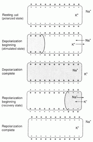 Figure 2-1. Depolarization and repolarization of a cardiac cell. |
Once a cell is stimulated, the membrane permeability changes. Potassium begins to leave the cell, increasing cell permeability to sodium. Sodium rushes into the cell, causing the inside of the cell to become more positive than negative (cell is depolarized). Muscle contraction follows depolarization. Depolarization and muscle contraction are not the same. Depolarization is an electrical event that results in muscle contraction, a mechanical event.
After depolarization, the cardiac cell begins to recover. The sodium-potassium pump is activated to actively transport sodium out of the cell and move potassium back into the cell. The inside of the cell becomes more negative than positive (cell is repolarized) and returns to its resting state.
Depolarization of one cardiac cell acts as a stimulus on adjacent cells and causes them to depolarize. Propagation of the electrical impulses from cell to cell produces an electric current that can be detected by skin electrodes and recorded as waves or deflections onto graph paper, called the ECG.
Electrical conduction system of the heart
The heart is supplied with an electrical conduction system that generates and conducts electrical impulses along specialized pathways to the atria and ventricles, causing them to contract (Figure 2-2). The system consists of the sinoatrial node (SA node), the interatrial tract (Bachmann’s bundle), the internodal tracts


Stay updated, free articles. Join our Telegram channel
Full access? Get Clinical Tree
