Electrodiagnostic Findings in Neuromuscular Disorders

Chapter 4 Electrodiagnostic Findings in Neuromuscular Disorders


Details of the electrodiagnostic findings in various neuromuscular disorders are outlined within the case studies in this book. The following is a brief summary of these findings in the most common neuromuscular disorders.


FOCAL MONONEUROPATHIES


Compression, traction, laceration, thermal, or chemical injury may damage one or more components of the peripheral nerves, including the myelin, axons, or supporting nerve structures (endoneurium, epineurium, and perineurium). The pathophysiologic responses to peripheral nerve injuries have a limited repertoire; that is demyelination, axon loss, or a combination of both.


Demyelinative Mononeuropathy


With focal injury to myelin, conduction along the affected nerve fiber is altered. This may result in slowing of conduction or conduction block along the nerve fibers or a combination of both.


1. Focal slowing. This is usually the result of widening of the nodes of Ranvier (paranodal demyelination). Focal slowing may be synchronized when demyelination affects all the large myelinated fibers equally. When the focal lesion is distal, there is prolongation of distal and proximal latencies while the proximal conduction velocity remains normal. If the focal lesion is between the distal and proximal stimulation sites, there is prolongation of proximal latency only resulting in slowing in proximal conduction velocity while the distal latency remains normal (Figure 4-1). With lesions manifesting as focal synchronized slowing, the CMAP amplitudes, durations, and areas remain normal and do not change significantly following proximal and distal stimulation. Desynchronized (differential) slowing occurs when conduction time is reduced at the lesion site along a variable number of the medium or small nerve fibers (average or slower conducting axons). Here, the CMAP is dispersed with prolonged duration on stimulations proximal to the lesion (Figure 4-2). The latency and conduction velocity along the injury site remain normal, since at least some of the fastest conducting axons are spared. When the largest axons are also affected, the dispersed CMAP with prolonged duration is also accompanied by slowing of distal latency (in distal lesions) or conduction velocity (in proximal lesions).

2. Conduction block. This is usually the result of focal loss of one or more myelin segment (segmental or internodal demyelination) which leads to interruption of action potential transmission. A nerve lesion manifesting with conduction block is best localized when it can be bracketed by two stimulation points, one distal to the site of injury and one proximal. In conduction block, stimulation distal to the lesion elicits a normal CMAP, whereas proximal stimulation elicits a response with reduced amplitude (partial conduction block) or absent response (complete conduction block) (Figure 4-3). The percentage drop in amplitude and area (amplitude or area decay) are calculated as follows:


image


image


There are several limitations to the definitive diagnosis of demyelinative conduction block:


Table 4-1 Electrodiagnosis of Conduction Block


Definite in Any Nerve*

Possible in Median, Ulnar, and Peroneal Nerves Only

* Caution should be taken in evaluating the tibial nerve, where stimulation at the knee can be submaximal, resulting in 50% or at times greater than 50% drop in amplitude and area, especially in overweight and very tall patients.


Axon-Loss Mononeuropathy


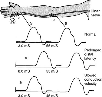
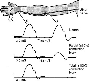
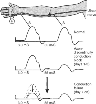
Following acute focal axonal damage, the distal nerve segment undergoes wallerian degeneration. However, early after axonal transaction, the distal axon remains excitable. Hence, stimulation distal to the lesion elicits a normal CMAP, whereas proximal stimulation elicits an absent response (complete conduction block) when the lesion is total and reduced CMAP amplitude (partial conduction block) when the lesion is incomplete (Figure 4-4). In an attempt to distinguish this pattern from a demyelinative conduction block, some refer to this pattern as an axonal noncontinuity, early axon loss, or axon discontinuity conduction block.


Wallerian degeneration of the axons distal to the nerve lesions is completed in 7–11 days. In the first 1–2 days, the distal CMAP and SNAP are normal. The distal CMAP amplitude then decreases and reaches its nadir in 5–6 days, while the distal SNAP amplitude lags slightly behind. It starts declining in amplitude after 4–5 days and reaches its nadir in 10–11 days (Figure 4-5). The earlier decline of the CMAP amplitude comparing to the SNAP amplitude following axon-loss nerve lesion is likely related to the early neuromuscular transmission failure that affects the recording of the CMAP amplitudes only. This is supported by the fact that MNAPs, recorded directly from nerve trunks, follow the time course of SNAPs.
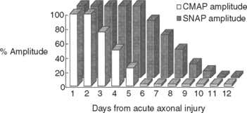


On motor NCS, a conduction block is present soon after axonal injury. However, as the distal axons undergo wallerian degeneration, this is replaced by unelicitable or low CMAP amplitudes with both distal and proximal stimulations corresponding to complete or partial motor axonal loss lesions respectively (see Figure 4-4). At this time, the distal CMAP amplitude is a reliable semiquantitative estimate of the amount of axonal loss in peripheral nerve lesions. In the chronic phases of partial axonal nerve lesions with reinnervation via collateral sprouting, the CMAP may improve to reach normal or near normal values giving a false indication of a milder degree of original axonal loss.


When the electrodiagnostic study is done early after an acute peripheral nerve lesion, it should be repeated at 10–11 days or later (or 5 days or later in purely motor nerves) in order to distinguish between conduction block caused by demyelination versus axonal loss, and to assess the extent of axon loss if present. Following this period of wallerian degeneration, stimulating the nerve below the lesion results in absent or reduced CMAP amplitude since degenerating axons would have lost their excitability. An absent or reduced CMAP amplitude from stimulation above or below the lesion indicates complete or partial axonal loss respectively. In demyelinating lesions, the distal CMAP remains unchanged with persistent conduction block across the lesion. In mixed lesions, the distal CMAP drops but remains significantly higher than the proximal implying both axon loss and segmental demyelination.


In partial axon-loss peripheral nerve lesions, the distal latencies and conduction velocities remain normal or are borderline. Selective loss of fast-conducting fibers associated with more than a 50% reduction in mean CMAP amplitude can slow conduction velocity to 80% of normal value because the velocity represents the remaining slow-conducting fibers. Motor conduction velocity may be occasionally slowed to 70% of normal value, when there is severe axonal loss with marked reduction of CMAP amplitude to less than 10% of normal.


Needle EMG is useful in assessing the progress of reinnervation of axon loss peripheral nerve lesions that may occur spontaneously or after nerve repair. Although collateral sprouting in partial axon loss lesions starts as early as 1–2 days after a nerve lesion, the early signs of reinnervation may first become evident on needle EMG one month later, but are usually definite by 2–3 months postinjury. MUAP morphology helps assessing the process of muscle fiber reinnervation that occurs following collateral sprouting and proximodistal regeneration of nerve fibers from the site of the injury. Collateral sprouting causes first an increased number of MUAP turns and phases followed by an increased duration and amplitude of MUAPs, while early proximodistal regeneration of nerve fibers in severe axon loss lesions often manifests by recording brief, small, and highly polyphasic (nascent) MUAPs. MUAPs tend to become longer in duration and higher in amplitude with the passage of time due to improved synchrony of muscle fiber action potentials.


In contrast to demyelinating or mixed mononeuropathies, pure axon-loss peripheral nerve lesions cannot be localized by NCSs when studied after the completion of wallerian degeneration, since they are not associated with focal conduction slowing or block. The identification of conduction block in the early days of axonal loss is extremely helpful in localizing a peripheral nerve injury. Waiting for the completion of wallerian degeneration results in diffusely low or unevoked CMAPs (regardless of stimulation site), which does not allow for accurate localization of the injury site. Localizing a purely axon-loss mononeuropathy after the completion of wallerian degeneration depends on needle EMG, with principles that are similar to manual muscle strength testing used during the neurological examination. Typically, the needle EMG reveals neurogenic changes (fibrillation potentials, reduced MUAP recruitment, chronic neurogenic MUAP morphology changes) that are limited to muscles innervated by the injured nerve distal to the site of the lesion (Figure 4-6). In contrast, muscles innervated proximal to the lesion remain normal. Unfortunately, attempting to localize axon loss lesions solely by needle EMG has several shortcomings that may result in poor localization or, sometimes, mislocalization of the site of the nerve lesion. These include the following scenarios:


Stay updated, free articles. Join our Telegram channel

Aug 12, 2016 | Posted by in CARDIOLOGY | Comments Off on Electrodiagnostic Findings in Neuromuscular Disorders

Full access? Get Clinical Tree

Get Clinical Tree app for offline access