(1)
Ospedale Civile di Vigevano, Vigevano, Italy
Drug-Induced Changes
A number of cardioactive drugs can cause changes in the electrocardiogram (ECG), the antiarrhythmic agents in particular. The most important effects produced by these drugs are bradyarrhythmias, which have been discussed in detail in Chap. 5. In this chapter, we will examine some of the specific effects of individual drugs, with special emphasis on their proarrhythmic effects.
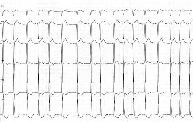
Figure 15.1 illustrates what is referred to as the digitalis effect. It is characterized by typical bowl-shaped depression of the ST segment, sometimes with shortening of the QT interval. These changes reflect the effect of digitalis (even at therapeutic blood levels) on the transmembrane action potential. Another commonly used drug, amiodarone, affects phase 3 of the cardiac action potential, thereby prolonging the QT interval. This is especially common during long-term treatment. The QT interval is sometimes markedly prolonged (Fig. 15.2), and this increases the risk of severe arrhythmias like torsades de pointes (Fig. 15.3). QT-interval prolongation is the most classic proarrhythmic effect of the antiarrhythmic drugs (see also Chap. 6). Iatrogenic arrhythmias related to a prolonged QT interval are also caused by sotalol, a beta-blocker whose effects on phase 3 of the action potential are just as powerful as those of amiodarone (Fig. 15.4). Finally, the Class IC antiarrhythmics, flecainide and propafenone, can produce significant ECG changes. Figure 6.31 shows an ECG recorded during flecainide therapy, which documents the presence of atrial flutter with 1:1 atrioventricular conduction and aberrant QRS morphology. As for propafenone, it causes changes that resemble those of the Brugada syndrome (see Chap. 9): RBBB and ST-segment elevation in leads V1 and V2 (Fig. 15.5). (In this patient, the presence of the genetic syndrome had been excluded.)


