ELASTIC PROPERTIES OF THE RESPIRATORY SYSTEM

3


ELASTIC PROPERTIES OF THE RESPIRATORY SYSTEM


Introduction

One of the properties of the respiratory system most often changed by disease is the ease with which it can be expanded and contracted in breathing.


The lungs and the chest wall that surrounds them are elastic structures, that is, they return to their original shape if a force that is distorting them is removed. The lungs have no muscles capable of changing their shape. The chest, on the other hand, is well supplied with internal and external intercostal muscles which can change its shape, and is separated from the abdomen by the most important inspiratory muscle, the diaphragm. Despite being closely pressed against it the lungs are not attached to the chest wall: there is a space of a few millimetres filled with a slippery plasma-like fluid. Because of this separation we can conveniently deal with the properties of the lungs and chest wall separately, but bearing in mind that in life they work together.


We can use a simple model to describe how changes in the volume of the elastic lungs are brought about by the changes in pressure around them.


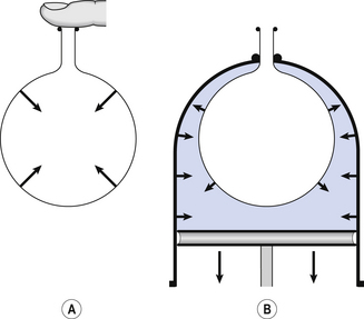
The most commonly used model of lung inflation is a toy balloon. Many of the principles that follow can be demonstrated by this model. For example, if you inflate a balloon and prevent the air escaping by blocking the neck with your finger (Fig. 3.1A), the elastic recoil of the balloon will be proportional to its elastance (1/compliance, see below) and will produce a recoil pressure. The pressure inside the balloon will be the same throughout if no flow is taking place into or out of the balloon. These observations demonstrate important principles of lung function.


An even more physiological model of the respiratory system can be made by suspending a balloon in a jar with a piston at its base, like a large syringe (Fig. 3.1B). In this case the balloon represents the lungs, the jar represents the chest wall and the piston represents the diaphragm. Lowering the piston reduces the pressure round the balloon (intrapleural pressure) and causes it to inhale.


Intrapleural pressure (Ppl)


For an object to be stretched or in some other way distorted it must be subjected to a force. In the case of a three-dimensional object this force may be pressure. In our simple model of breathing (Fig. 3.1A), inspiration would be inflation of the balloon and expiration deflation. The pressure that brings about inflation in Figure 3.1A would be applied to the inside. There is a pressure gradient from inside the balloon to outside. The other, more complicated, way for us to inflate the balloon would be to reduce the pressure outside it using the jar and plunger (Fig. 3.1B): again there is a pressure gradient from inside to outside the balloon, and this is the way we inflate our lungs.


Even when we are completely relaxed, at the end of an expiration with no contraction of the respiratory muscles there is a tension in the thorax between the lungs, whose elasticity is causing them to collapse, and the chest wall whose elasticity is causing it to spring outward. These two structures are ‘locked together’ by the intrapleural fluid in the intrapleural space. Because it is a fluid and therefore not compressible or expandable, and the intrapleural space is airtight, the lungs are as firmly pressed to the chest wall as a suction cup attached to a window.


The tension between the lungs trying to collapse and the chest wall trying to spring out is most clearly seen in surgery, when the sternum is split to allow the surgeon to get at the heart, for example: the lungs collapse and the ribs spring out.


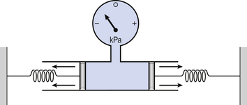
Another way of visualizing what is happening in the space between the lungs and chest wall is to imagine a syringe with two plungers being pulled in opposite directions (Fig. 3.2).


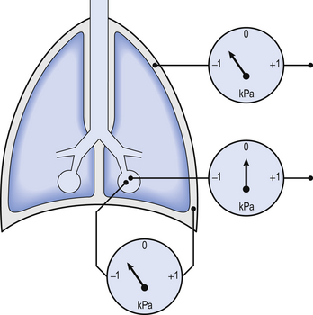
You can see from such a model that intrapleural pressure is negative with respect to atmospheric pressure. What is not immediately obvious is that intrapleural pressure is also negative with respect to air pressure within the alveoli, because the alveoli are connected to the atmosphere by a system of open tubes, the bronchial tree (Fig. 3.3).


This means a hole made between either the atmosphere or the alveoli and the intrapleural space will allow the pressure surrounding the lung to rise and the lung to collapse: this dangerous condition is called a pneumothorax.


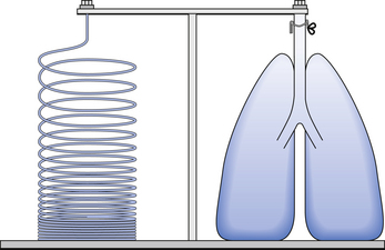
Because the lungs are to some extent suspended from the trachea and rest on the diaphragm, they behave like a child’s ‘slinky’ (a very soft spring), held at one end and supported from underneath. Gravity causes the spring or lungs to slump under their own weight (Fig. 3.4).


Because of this effect the chest behaves as if it is filled with a liquid of the average density of the lungs.


As you descend below the surface of any liquid gravity causes pressure to increase at a rate dependent on the density of the liquid. The intrapleural pressure therefore increases (in fact becomes less negative) as you move from the apex to the base of the lung. At the end of expiration the pressure is about –0.8 kPa at the top of the lungs and –0.2 kPa at the base. That this gradient of pressure depends on the effect of gravity on the contents of the thorax is clearly demonstrated by the fact that it reverses if the subject in which it is being measured stands on their head.


At any instant the negative pressures that surround the lung expand it to a given volume. If those pressures did not change lung volume would not change and we would not breathe. We cause our lungs to breathe by changing the negative pressure around them by making the diaphragm contract and, by acting like the plunger of a syringe, draw air into the chest.


Intrapleural pressure can be measured by inserting a hollow needle between the ribs into the intrapleural space. It is not easy to obtain volunteers to undergo this procedure, and as we are usually more interested in changes in intrapleural pressure than their absolute values, we frequently measure changes in pressure in the oesophagus, which forms a very flexible tube running through the thorax. Because the oesophagus is so flexible the changes of pressure within it closely follow changes in intrapleural pressure.


Static lung compliance (CL)


We have seen that the lungs are elastic structures, i.e. they return to their original shape and size when distorting forces are removed. These distorting forces are usually those of intrapleural pressure, which becomes more negative to bring about inspiration and then becomes less negative, and the elasticity of the lungs leads to quiet expiration. This elasticity is a measure of how easily the lungs can be stretched and is conventionally expressed as compliance, the reciprocal of elastance. This ‘stretchiness’ of the lungs can be measured under static conditions, i.e. by measuring pressure and volume when there are no breathing movements taking place, or as dynamic compliance during breathing (see below).


The effect of disease


The compliance of the lungs is changed by most lung diseases. Such changes have a detrimental effect on lung function and increase the work of breathing. It seems that healthy lungs are at optimal compliance, and an increase or decrease from this norm is for the worse. For example, in lung fibrosis the lungs are stiffened by the laying down of collagen and fibrin bundles, so that compliance is reduced. In emphysema the parenchyma of the lungs is destroyed, there is less elastic recoil, and compliance is therefore increased. In infant respiratory distress syndrome it is the nature of the liquid lining the lungs that is at fault (see below), and this also reduces lung compliance. The origin of these changes becomes clear if we consider the two physical systems that contribute to the elasticity of the lungs and hence their compliance. One originates in the elasticity of lung tissues, the other depends on the nature of the liquid lining of the alveoli.


The physical basis of lung compliance


The elastic properties of the lungs, and hence their compliance, depends almost equally on the elastic properties of their tissues and on the elastic properties of their liquid lining.


Case 3.1   Elastic properties of the respiratory system: 2


Causes of RDS of the newborn


The principal cause of RDS of the newborn is a deficiency of lung surfactant related to prematurity, although the disease is also related to the general immaturity of a premature baby’s respiratory system. The more premature an infant, the more likely it is to develop RDS.


The type II pneumocytes that produce surfactant develop at about 24 weeks’ gestation, although most fetuses do not start producing large amounts of surfactant until about 34 weeks (babies are born, on average, at 40 weeks). Premature babies also have smaller lungs and alveoli than full-term babies. Remember the law of Laplace, which states that:


image


where P is the pressure inside a bubble, T is the surface tension and R is the radius. In the alveoli of premature infants T is greater than normal because of the lack of surfactant, and R is less than normal because the premature infant has smaller alveoli. For both these reasons, a high pressure (P) is needed to keep the alveoli open. This means that the lungs tend to collapse during expiration, and the effort of inspiration is very much increased. Furthermore, the lack of surfactant means that fluid tends to be drawn from the blood into the alveoli, which therefore become oedematous. All these things mean that the dynamic compliance of the lungs is very much decreased.


Because it has not developed fully the compliance of a premature infant’s chest wall is high, and this means that respiratory effort causes indrawing of the chest wall. Grunting in infants is thought to be an effort to increase airway pressure during expiration, which would tend to reduce airway collapse. Blood still flows through the collapsed areas of the lungs but remains deoxygenated. Without treatment, the baby’s respiratory distress would become worse and would eventually lead to respiratory failure as the baby became increasingly exhausted and hypoxic by the effort of breathing.


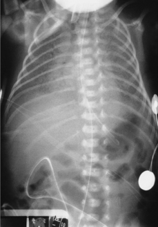
The large number of collapsed alveoli lead to a characteristic chest X-ray appearance. Baby Aldridge’s chest X-ray is shown in Figure 3.5. The collapsed alveoli give the chest X-ray a ‘ground-glass’ appearance. Against this are visible air bronchograms which are the shadows of the gas within the bronchi. These are only visible on a chest X-ray if the lung tissue around them is unusually dense.


The lungs of infants who have died from RDS have a characteristic appearance under the microscope. The alveoli are collapsed and they and the respiratory bronchioles appear to be lined with a membrane. This membrane is made up of a proteinaceous exudates from the airways and can, in fact, be caused by many types of lung injury, not only RDS. Because of its appearance with commonly used histological stains, the membrane is usually described as hyaline, meaning nearly transparent. For this reason, RDS is also called hyaline membrane disease.


Jun 18, 2016 | Posted by in RESPIRATORY | Comments Off on ELASTIC PROPERTIES OF THE RESPIRATORY SYSTEM

Full access? Get Clinical Tree

Get Clinical Tree app for offline access