Fig. 6.1
EKG at rest while the patient was asymptomatic
Echocardiography (Not Shown)
Normal both atria size (LA diameter M-mode = 3.6 cm; area 4c = 18 cm2, RA area 4c = 16 cm2). Normal size and wall thickness of the left ventricle (iLVEDV 42 ml/m2), which shows a preserved systolic function (ejection fraction with Simpson’s method 0.67). Normal size and global function of the right ventricle (TAPSE 23 mm).
Normal morphology of the cardiac valves; mild tricuspid regurgitation and normal systolic pressure gradient (PASP = 25 mmHg). Normal size of the aortic root (24 mm) and of the proximal ascending aorta (28 mm); normal size of the aortic arch (26 mm) and the descending thoracic aorta (22 mm) in the explored segments too. The inferior vena cava has also a normal size (12 mm) and physiological collapsing during inspiration. Normal diastolic pattern without increased filling pressure (E/A 0.9, E/E’ 6, E dec time 210 m/s).
Absence of pericardial effusion.
Conclusion: normal echocardiographic findings for a 38-year-old man.
Routine Laboratory Tests
Complete blood count: normal.
Cholesterol (total, HDL, LDL) and TG: normal.
Hepatic function (GOT, GPT, γ-GT, ALP, total bilirubin, direct and indirect): normal.
Thyroid function (TSH, FT3, FT4): normal.
Renal function (creatinine, BUN): normal.
Electrolytes (Na + , K + , Ca ++ , Mg ++ , Cl − ): normal.
Fasting blood glucose: 102 m/dl (5.67 mmol/L).
Troponin I-hs: 0.012 ng/ml (n.v < 0.055 ng/ml)
Inflammation index: VES 35 mm/h (n.v < 27 mm/h), CRP 0.2 mg/dl (n.v <0.6 ng/ml)
D-dimers: 150 ng/mL (n.v. < 280 ng/mL)
The echocardiographic findings are completely normal, so we can rule out cardiomyopathies and any severe valvular disease as previously supposed.
Similarly, the aortic size is normal, and there are no signs of acute aortic dissection (intimal flap) or intramural hematoma. Furthermore, the biomarkers are normal (D-dimer and troponin), and the symptoms referred by the patients are not typical of acute aortic syndrome. Moreover, he didn’t present any cardiovascular risk factor, with the exception of the smoking habit. He did not have any familial history of arterial disease.
The EKG recorded while the patient was asymptomatic showed ST-segment elevation in precordial leads (V2–V6), with positive T wave and a concave pattern, without any reciprocal ST-segment changes in the other leads.
The Most Common Causes of ST-Segment Elevation in a Young Man
Acute coronary syndrome with ST-segment elevation (ACS-STEMI) with or without possible coronary spasm
Myocarditis/pericarditis
Early repolarization pattern
Acute pulmonary embolism
Hyperkalemia
Brugada syndrome
Acute aortic dissection
The first hypothesis we have to exclude is ACS-STEMI. Even though there was an ST-segment elevation in more than one precordial lead, it should be ≥0.25 mV when measured at the J point in a man under 40 years. Moreover, the upward concavity is not typical of STEMI; the limb leads showed an essentially normal repolarization pattern, and there were no reciprocal EKG changes. By considering also normal echocardiography and laboratory results and the clinical history (previous recent coronary angiography was completely normal), we excluded the ACS hypothesis.
The EKG pattern in possible coronary spasms is also different because in addition to the ST-segment modification the R wave may become taller and the S wave may decrease in amplitude or disappear in the leads in which the ST elevation is observed (see Chap. 1).
The repolarization abnormalities in our patient were not typical of an acute pulmonary embolism, which is usually characterized by sinus tachycardia, or other atrial arrhythmias, complete or incomplete right bundle branch block, “pulmonary P wave,” ST-segment abnormalities, and/or inverted T wave in right precordial leads. Clinical signs and symptoms and the normal D-dimer level highly contribute to exclude pulmonary embolism.
There were no peaked T waves (“tended”) in right precordial leads as we could expect during hyperkalemia; moreover, the laboratory tests ruled out any electrolyte abnormality.
The EKG pattern was also not typical of Brugada type 1 pattern (ST-segment elevation ≥2 mm descending with an upward convexity, inverted T wave in at least one right precordial lead); the clinical history was also negative for syncope or tachyarrhythmic episodes.
Acute pericarditis could be more likely, especially according to chest pain, early recurrence, and its characteristics. Usually in this pathology, the ST-segment elevation is diffuse in all the leads and little pronounced; QRS complex generally shows a reduction in amplitude, and PR can be depressed in many leads. In myocarditis, the most common EKG finding is diffuse T wave inversion, whereas ST-segment displacement is less common. The absence of flu during the days/weeks before, the normal values of the logistic index at the laboratory tests together with the absence of pericardial effusion or wall motion abnormalities made those hypotheses unlikely.
Final Diagnosis
According to the clinical history, echocardiographic findings, and laboratory tests, we concluded that the EKG abnormalities were due to an early repolarization pattern.
An exercise testing was performed showing a complete normalization of the ST-segment elevation during exercise with a slow and progressive return during recovery (Fig. 6.2). That confirmed our diagnostic hypothesis.
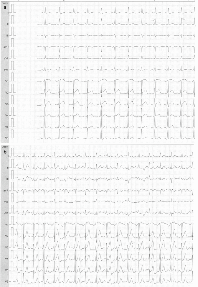
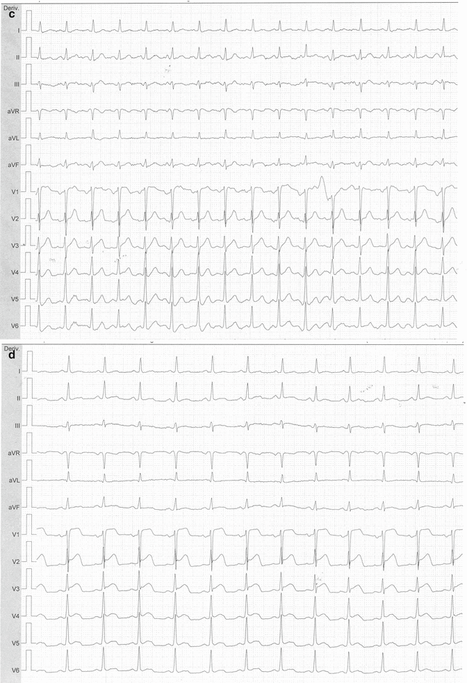
Fig. 6.2
EKG recorded during exercise testing showing the ER pattern at rest (a), normalization of the ST-segment elevation during exercise (b); slow return to the baseline and then progressive return of the ER pattern (c, d)
This EKG pattern is more frequent in black men and does not require any pharmacological treatment.
There is no common agreement if different ER patterns (commonly separated in benign and malignant forms, see below) are associated with a higher risk of SCD. Nevertheless, our patient’s EKG showed a benign pattern because the ST-segment elevation was present only in precordial leads, with upper concavity, and the T waves were large and positive. There was a J wave in some leads (V4–V5–V6), but it was little and did not show significant modifications. The complete repolarization pattern remained stable during the hospitalization period without significant changes at rest. Moreover, the patient did not present proarrhythmic conditions and did not refer familiar history of SCD. So he could be safely discharged.
However, for completing diagnostic evaluation, we decided to perform an EGDS, which showed a grade B gastroesophageal reflux disease (Los Angeles classification) with some mucosal breaks that did not extend between the tops of the two mucosal folds. Proton pump inhibitor therapy has been prescribed, and a gradual decrease of the chest pain was observed during the following days. Finally, the patient was discharged suggesting an additional gastroenterological reassessment in the next weeks after full therapy.
< div class='tao-gold-member'>
Only gold members can continue reading. Log In or Register to continue
Stay updated, free articles. Join our Telegram channel

Full access? Get Clinical Tree


