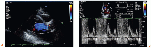
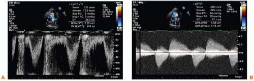
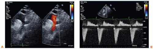
A 67-year-old man with a history of hypertension presents with 3 months of increasing dyspnea, orthopnea, and lower extremity edema. His transthoracic echocardiogram is shown in Figures 56-1, 56-2 and 56-3.
Figure 56-1A,B.
Figure 56-2A,B.
Figure 56-3A,B.
QUESTION 1. All of the following would support a diagnosis of severe aortic regurgitation (AR) except:
A. Jet width > 65% left ventricular outflow tract (LVOT)
ANSWER 1: C. A regurgitant volume > equal to 60 mL per beat is highly suggestive of severe AR and not 50 mL per beat (Table 56-1).
TABLE 56-1. Qualitative and Quantitative Parameters Useful in Grading Aortic Regurgitation Severity
Mild
Moderate
Severe
Structural parameters
LA size
Normal
Normal or dilated
Usually dilated
Aortic leaflets
Normal or abnormal
Normal or abnormal
Abnormal/flail or wide coaptation defect
Doppler parameters
Jet width in LVOT -Color Flow
Small in central jets
Intermediate
Large in central jets; variable in eccentric jets
Jet density-CW
Jet deceleration rate – CW (PHT, ms)
Incomplete or faint slow > 500
Dense Medium 500-200
Dense Steep < 200
Diastolic flow reversal in descending aorta -PW
Brief, early diastolic reversal
Intermediate
Prominent holodiastolic reversal
Quantitative parameters
VC width, cm
< 0.3
0.3-0.60
> 0.6
Jet width/LVOT width, %
< 25
25-45
46-64
≥ 65
Jet CSA/LVOT CSA, %
<5
5-20
21-59
≥ 60
R Vol, ml/beat
< 30
30-44
45-59
≥ 60
RF, %
< 30
30-39
40-49
≥ 50
EROA, cm2
< 0.10
0.10-0.19
0.20-0.29
≥ 0.30
Reprinted from Zoghbi WA, Enriquez-Sarano M, Foster E, et al. Recommendations for evaluation of the severity of native valvular regurgitation with two-dimensional and Doppler echocardiography. J Am Soc Echocardiogr. 2003; 16:777-802, with permission from Elsevier.
Only gold members can continue reading. Log In or Register to continue