Figure 38.2 Pulse wave Doppler recording with the sample volume in the orifice of hypertrophied muscle band (arrow), the systolic and diastolic signals are well seen (a). The peak early diastolic velocity is 156 cm/sec and estimated gradient between two right ventricle chambers is 10 mmHg (b).
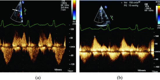
Figure 38.3 A hypertrophied muscle bundle divide the right ventricle cavity into a proximal and a distal chamber. The muscle bundle (arrow) run between an area located in the ventricular septum, beneath the level of the septal leaflet of the tricuspid valve, and the anterior wall of the right ventricle demonstrated by transesophageal echocardiography in the longitudinal plane (a). A right to left shunt (arrow) through the patent foramen oval is indicated by transesophageal echocardiography with color Doppler (b). LA, left atrium; LV, left ventricle; RA, right atrium; RV, right ventricle.
Stay updated, free articles. Join our Telegram channel

Full access? Get Clinical Tree


