Doppler Profiles and Assessment of Diastolic Function

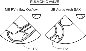
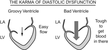
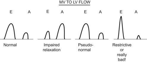
Chapter 8


Doppler Profiles and Assessment of Diastolic Function


Christopher J. Gallagher and John C. Sciarra


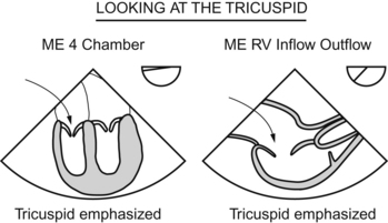
Tricuspid Valve and Right Ventricular Inflow


The big Mammas of the valve world are the mitral and aortic, with whole lectures dedicated to each one individually. The tricuspid and pulmonic valves usually get lumped together, like third-class steerage passengers on the Titanic. It’s a safe bet that you should put your efforts into the mitral and aortic valves. But, let’s soldier on through the tricuspid valve.


The tricuspid valve has (duh) three cusps. Set in a (normally) low-pressure system, the tricuspid doesn’t “have to” function perfectly. You will normally see some regurg here. Think about it; sometimes the tricuspid valve is removed and not even replaced, and the heart continues to function. Try that with the aortic valve!


You get a dandy view of the tricuspid in the “easiest” view to get, the ME four-chamber. The ME RV inflow–outflow view also gives you a shot at the tricuspid.


Focusing your Doppler on the tricuspid valve will tell you just how severe the tricuspid regurg is. The regurg is severe if:


Also, if the hepatic vein flow profile shows systolic flow reversal, that also indicates severe tricuspid regurgitation. If you think about it, that makes sense. When the heart contracts, if the tricuspid valve doesn’t work, the blood flows back into the right atrium and just keeps on flowing backward, backward, backward, all the way back into the inferior vena cava and back further into the hepatic vein (which feeds into the inferior vena cava).


image


Pulmonary Valve and Right Ventricular Outflow


Look at your model of the heart again. The pulmonary valve is the farthest away valve in the heart. No surprise, then, that you sometimes have a hard time getting a good look at it. And getting a good Doppler study through it can be a real pain.


Fortunately, a kind Providence has given us a few good views of the pulmonic valve. The ME RV inflow–outflow view works. If you get that aortic valve in a good en face view (the Mercedes Benz sign), then you will get a 90-degree view of the pulmonic valve.


Another view (not quite as easy to get) is the UE aortic arch SAX view. This view gives you a better chance at getting a Doppler shot down the pipe of the pulmonic valve.


image


When you Doppler-ifize the pulmonic valve, you will see some regurg, especially if there is a PA catheter straddling the valve. As with other valves, you can get an impression of the degree of regurg by looking at the size and depth of the regurgitant jet. A big jet means a lot of regurg, a little jet means a little regurg. (Aren’t you glad you went to school for years and years to be able to figure out such complex stuff?)


Since the pulmonic valve lies far afield from the echo probe, getting more quantitative than that just ain’t in the works.


When you see PR, it’s worth thinking about what might be causing it. As with other valves, a poorly functioning valve (endocarditis, carcinoid syndrome, congenital defect) may account for the blood flowing backward. Also, highpressure “downstream” of the valve (pulmonary hypertension, pulmonary embolus) may “overwhelm” a normal valve and cause regurgitation.


Mitral Valve and Left Ventricular Inflow


How the blood flows into the ventricle tells a lot about the ventricle’s function during diastole. If the heart is healthy, springy, and not stiff, then the blood will flow in easily. If the heart is sick, stiff, and nonresilient, then the blood will have to “work hard” to fill the ventricle.


image


Judging by the puzzled looks in the diastolic dysfunction lectures, it was evident to me that this whole idea is a little hard to absorb. For some reason, the idea of systolic dysfunction is easy to grasp:


The pump doesn’t work, but the concept of diastolic dysfunction is tough.


The loading of the pump doesn’t work.


The world would be a nice place if the mitral inflow patterns were a simple


But noooooooooooooooooooooooooooooooooooooooooooo! Life is not so. Understanding these confusing patterns is the whole crux of understanding diastolic dysfunction.


First, I’ll blast through the patterns, then I’ll go back and try to drag you through the reasoning. PLOW THROUGH THIS STUFF SLOWLY, AND LOOK FOR THE EXPLANATION IN A FEW DIFFERENT BOOKS, FOR DIASTOLIC DYSFUNCTION IS A TOUGHIE.


Now, the reasoning behind the patterns. (The first part is easy—the second is a bit sticky.)


Side note: to acquire the E/A ratio, place the pulse wave circle thing at the tip of the mitral leaflets. For the pulomonary flow go way to the top of the left atrium and put the PW there. You may have to ask a professional for help with this.


image


Stay updated, free articles. Join our Telegram channel

Jun 4, 2016 | Posted by in CARDIOLOGY | Comments Off on Doppler Profiles and Assessment of Diastolic Function

Full access? Get Clinical Tree

Get Clinical Tree app for offline access