Fig. 27.1
Results of a renal colour-Doppler ultrasonography showing renal vascularization (a). RI measurement using pulsed-wave Doppler (b)
Stage | Quality of renal perfusion by colour-Doppler |
|---|---|
0 | Unidentifiable vessels |
1 | Few vessels in the vicinity of the hilum |
2 | Hilar and interlobar vessels in most of the renal parenchyma |
3 | Renal vessels identifiable until the arcuate arteries in the entire field of view |
To characterize the intrarenal Doppler waveform, most investigators have used the resistive index (RI) so-called Pourcelot index (Fig. 27.1b). Three to five reproducible waveforms are obtained, and RIs from these waveforms are averaged to compute the mean RI for each kidney. This easily calculated parameter is defined as follows:
![$$ \mathrm{R}\mathrm{I}=\left[\mathrm{peak}\;\mathrm{systolic}\;\mathrm{shift}-\mathrm{minimum}\;\mathrm{diastolic}\;\mathrm{shift}\right]/\mathrm{peak}\;\mathrm{systolic}\;\mathrm{shift} $$](/wp-content/uploads/2017/04/A320045_1_En_27_Chapter_Equa.gif)
RI can theoretically range from 0 to 1. It is normally lower than 0.70. In several studies, mean RI (±SD) in healthy subjects ranged from 0.58 (±0.05) to 0.64 (±0.04) [23, 24]. The normal RI range is however age dependent. Thus, RI values greater than 0.70 have been described in healthy children younger than 4 years [25] and in individuals older than 60 years and considered healthy [26]. When the RI is measured for both kidneys, the side-to-side difference is usually less than 5 % [27].
Since RI depends in part on the minimum diastolic shift, it may be influenced by the heart rate [28]. According to observations performed by Mostbeck and colleagues regarding RI changes as consequences of heart rate variations, a formula has been developed to correct the RI value for heart rate: [corrected RI = observed RI −0.0026 × (80-heart rate)] [28]. However, the influence of heart rate per se on RI remains unclear, and this formula is not used in most of the studies evaluating interest of RI [29, 30]. Arrhythmias and especially atrial fibrillation may impact renal RI measurement and interpretation.
Renal RI is a simple and non-invasive tool easy to use at the patient’s bedside. Feasibility of the measure has been shown to be good, even in the settings of critically ill patients, and a recent study suggested a short (half-day) training session might allow inexperienced operators to perform renal Doppler [21]. Interobserver reproducibility of RI measurement by senior radiologist or senior intensivist is considered excellent [14, 31]. In critically ill patients, the interobserver reproducibility between senior and inexperienced operator is good, and measures seem accurate although associated with a lack of precision [21].
Renal pulsatility index is another parameter derived from renal blood flow velocities and is calculated as follows:
![$$ \mathrm{PI}=\left[\mathrm{peak}\;\mathrm{systolic}\;\mathrm{velocity}-\mathrm{minimum}\;\mathrm{diastolic}\;\mathrm{velocity}\right]/\mathrm{mean}\;\mathrm{velocity} $$](/wp-content/uploads/2017/04/A320045_1_En_27_Chapter_Equb.gif)
Both RI and pulsatility index are however closely correlated (r = 0.92; P < 0.001) [13], and RI might be more adapted to the study of high-resistance vascular territories explaining why most authors report RI. Additionally, pulsatility index has been shown to be subject to wider variations than RI (reproducibility 9–22 % vs. 4–7 %) [32].
Contrast-enhanced US is believed to allow an accurate quantification of regional and global renal blood flow [19]. This technique relies on the intravascular injection of specific contrast agents that create a signal of high echogenicity, thus allowing macro- and microvascular structure visualization when using specific imaging techniques. These specific contrast agents consist in gas-filled microbubbles that oscillate in response to US waves, therefore creating a non-linear signal of high echogenicity [33]. It has been validated in humans to evaluate coronary blood flow [34], and its safety has been largely documented in this context [35]. When adding this technique to recently developed softwares, this technique is believed to allow an accurate quantification of regional blood flow, such as renal blood flow [19]. A recent study has confirmed feasibility of this technique in cardiac surgery patients, and additional studies are ongoing and should help in more clearly assessing input of this technique [36].
27.3 Physiological Significance of the Renal Resistive Index
Although renal Doppler has been the focus of many studies, the physiological significance of the RI remains debated. As suggested by its name, the RI was initially considered an indicator of renal vascular resistance and blood flow [7]. However, experimental and clinical studies have suggested correlation of RI with vascular resistance and blood flow to be weak [16, 17]. In fact, several hemodynamic and physiological factors influence the intrarenal arterial Doppler waveform patterns and, therefore, the RI value (Table 27.2).
Physiological factors | Vascular compliance (arterial stiffness) |
Vascular resistances | |
Pulse pressure | |
Renal blood flow | |
Heart rate | |
Oxygen and carbon dioxide levels | |
Age | |
Pathological factors | Interstitial pressure |
Ureteral pressure | |
Intra-abdominal pressure | |
Mechanical ventilation with positive pressure |
Several studies established that vascular compliance is crucial to the interpretation of RI values [16, 17, 29]. In an in vitro study, the relationship between vascular resistance and RI was linear only when vascular compliance was normal and progressively disappeared when vascular compliance decreased [17]. This was confirmed in studies in ex vivo rabbit kidney models [16, 29]. In addition, the observed RI changes in response to pharmacologically induced changes in renal vascular resistance were modest [29]. In this model, large, non-physiological, pharmacologically induced changes in renal vascular resistances translated into slight changes in RI (RI changes of 0.047 IU (+/−0.008) per logarithmic increase in renal resistances) [29]. In the same model, changes in pulse pressure index [(systolic pressure – diastolic pressure/systolic pressure)] had direct and dramatic effects on RI values [29]. Clinical data supporting influence of impaired vascular compliance in determination of RI are scarce. However, a recent longitudinal cohort study performed in renal allograft recipients gave interesting information as regards to this relationship [10]. Hence, the main factor associated with elevated RI in renal transplant recipient was found to be characteristics of both donor and receiver rather than renal transplant function or outcome [10]. In addition to these factors, both oxygen and carbon dioxide levels can affect RI. Several studies have demonstrated RI, or pulsatility index or RI, to vary according to PaO2 and PaCO2 levels [37–39]. These studies performed in healthy subject, patients with chronic obstructive respiratory disease, renal transplant recipients or patients with acute respiratory distress syndrome suggest that hypoxemia and hypercapnia may increase RI [37–40].
As stated above, RI has been shown to increase with age [26]. Age-related arterial stiffening may explain this phenomenon mainly by increasing central pulse pressure (i.e. increased central arterial stiffness) and maybe by decreasing intrarenal arterial compliance (i.e. increased intrarenal arterial stiffness) [41]. Similarly, elevated RI observed in several pathological states such as diabetes mellitus and hypertension may also be related to influence these diseases on arterial stiffness [42, 43]. It must be pointed out that renovascular disease is common in older subjects and might greatly influence RI. Some authors suggested an increased peak systolic velocity (more than 1 m per second) to be highly sensitive and specific to detect renal artery stenosis [42].
Besides vascular and hemodynamic factors, kidney interstitial pressure has been shown to be associated with RI in ex vivo studies [16]. An increase in interstitial pressure reduces the transmural pressure of renal arterioles, thereby diminishing arterial distensibility and, consequently, decreasing overall flow and vascular compliance. Similarly, intra-abdominal pressure may affect RI via the same mechanisms. Thus, incremental changes in intra-abdominal pressure correlated linearly with RI in a porcine model [44], and reduction in intra-abdominal pressure with paracentesis was followed by a decrease in RI in cirrhotic patients with tense ascites [45]. Finally, ureteral pressure, likely acting via interstitial pressure, also affects RI [6].
27.4 Doppler-Based Renal Resistive Index and Hypertension
As stated in the previous paragraph, the Doppler-based renal RI is an integrated index of arterial compliance, resistance, pulsatility and perfusion [17, 29]. In addition, RI has been demonstrated to be correlated with renal arteriosclerosis in numerous studies [10, 46]. Potential interest of Doppler-based renal RI has therefore been evaluated in assessing renal function or risk of end-organ damage in patients with hypertension or diabetes mellitus.
In patients with never-treated essential hypertension, a weak but significant correlation was observed between RI and both intima-media thickness (r = 0.25; P < 0.0001) and ambulatory arterial stiffness index (r = 0.27; P < 0.0001) suggesting a correlation between RI and early organ damage in hypertensive patients [47]. Other studies found similar results [48, 49]. Additionally, elevated RI was found to be associated with other targeted organ damage, namely, left ventricular hypertrophy, aortic stiffness or albuminuria [48, 50–53]. Furthermore, elevated RI was also found to be associated with mild reduction in glomerular filtration rate [42, 52]. These findings suggest Doppler-based resistive index to be a potent predictor of preclinical organ damage and of preclinical arteriosclerosis and therefore a predictor of cardiovascular risk profile [49].
Subsequently, several studies evaluated relationship between Doppler-based RI and outcome. Hence, Doi and colleagues demonstrated RI higher than 0.7 to be associated with cardiovascular events, end-stage renal disease or death in patients with hypertension (Fig. 27.2) [54]. Similarly, Pearce and colleagues found Doppler-based end-diastolic velocities to be associated with cardiovascular event or death in elderly patients [55]. In this study, however, only end-diastolic velocities (and not RI) were found to be associated with these events [55]. Although these studies give interesting insight as regards the potential interest of Doppler-based RI, no study to date gave an adequate cut-off or an estimation of RI performance in predicting adverse events. Additional studies in this field are therefore needed before implementing Doppler-based RI in assessing prognosis of hypertensive patients.
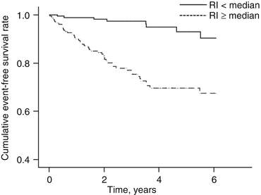
Fig. 27.2
Probability of survival free of cardiovascular and renal event (all-cause death, stroke, myocardial infarction, congestive heart failure requiring hospitalization, aortic dissection or end-stage renal failure) in patients with hypertension and normal (RI <0.65 or <0.68 in patients of male gender and female gender, respectively) or elevated RI (Reproduced with permission from Doi et al. [54])
Interestingly, a preliminary report suggested Doppler-based RI to decrease accordingly to urine albumin excretion in patients receiving ACE inhibitors [56]. Similarly, RI was found to be decreased in parallel to blood pressure and urine albumin excretion following renal denervation [57]. Last, some authors found Doppler-based RI to be a predictor of renal outcome following renal revascularization in patients with unilateral renal artery stenosis [58]. In this study, however, blood pressure outcome was not associated with RI [58]. Although some confirmation studies are needed in this field, these findings might suggest Doppler-based RI to be of help in choosing, adapting and optimizing treatment in hypertensive patients.
27.5 Doppler-Based Renal Resistive Index in Selected Renal Diseases
Studies started in 1990 have evaluated the performance of RI measurement as a tool for assessing renal perfusion [59]. A preliminary study evaluated correlations between RI values and renal biopsy findings in patients with renal diseases requiring renal biopsy [60]. In this preliminary work, patients with interstitial, tubular, or vascular nephritis had markedly increased RI values (mean, 0.73–0.87), whereas patients with isolated glomerular disease had normal RI values (mean, 0.58) [60]. However, subsequent studies did not confirm these findings. Thus, in non-transplanted patients, RI failed to distinguish among five groups of predefined renal parenchymal diseases [61]. Similarly, several studies found that RI contributed to predict recovery from haemolytic and uremic syndrome [5], to detect renal involvement in systemic sclerosis [62], to predict renal outcomes in lupus nephritis [63] or to evaluate the progression of chronic renal diseases [64]. However, most of these studies were preliminary studies, and validation studies are still lacking in these different settings.
Several studies have evaluated the usefulness of RI measurement for assessing the risk of renal dysfunction after renal transplantation [4, 9, 10, 65]. Preliminary reports suggested that RI elevation might be highly specific of acute rejection [4, 8]. However, these findings were invalidated by subsequent studies [9, 10]. Nevertheless, several studies found that RI elevation predicted poor long-term outcomes [9, 10, 66] and accurately predicted early vascular and non-vascular renal complications (e.g., acute kidney injury, rejection and obstructive kidney disease) [67]. In a recent large prospective study, the main contributor to elevated RI was found to be characteristics of the donor or the recipient rather than renal histological features and that RI might be an integrative parameter of risk factors rather than the reflect of an acute renal events [10].
Lastly, renal Doppler has been evaluated for the early detection of AKI and for monitoring renal perfusion in critically ill patients [12]. Hence, in a preliminary study conducted in septic critically ill patients, RI measured at admission was significantly higher in patients who developed subsequently AKI [14]. This finding was recently confirmed in the post-operative setting of cardiopulmonary bypass [68]. Moreover, several studies have suggested that renal resistive index (RI) may help to discriminate between patients with transient AKI and those with persistent AKI [15, 69–71]. However, most of these studies are once again preliminary reports performed in limited population of patients, and confirmation studies are still lacking. In addition, a recent study reported discrepant results and a poor performance of RI in assessing renal dysfunction reversibility [72]. Lastly, significance of elevated RI in this population of patients remains questionable. Indeed, elevated RI might reflect renal interstitial oedema, decrease in renal perfusion, or reflect pre-existing preclinical renal arteriosclerosis and therefore a risk factor of persistent AKI.
27.6 Conclusion
Renal Doppler is a rapid, non-invasive and repeatable technique that may help in assessing renal preclinical dysfunction or vascular damages, in evaluating risk of subsequent renal dysfunction and evaluating severity of an acute kidney injury. This integrative parameter neither constitutes a substitute for renal biopsy nor provides reliable information on renal blood flow. Nevertheless, renal Doppler is a promising tool that might help in monitoring patients with hypertension, risk of cardiovascular diseases or acutely ill patients. Despite the promising preliminary results, validation studies are still awaited, and clinical relevancy of this technique is still uncertain. In addition, numerous factors have been shown to influence renal Doppler, each of them being also a potential confounder. Additional studies with larger population of unselected patients and pragmatic evaluation of the input of Doppler-based resistive index are therefore mandatory before implementing this test in clinical practice.
References
1.
Barozzi L, Valentino M, Santoro A et al (2007) Renal ultrasonography in critically ill patients. Crit Care Med 35:S198–S205. doi:10.1097/01.CCM.0000260631.62219.B9 CrossRefPubMed
Stay updated, free articles. Join our Telegram channel

Full access? Get Clinical Tree


