Fig. 19.1
(a) 2D echocardiogram and colour Doppler imaging of a high parasternal short-axis view with colour Doppler showing the aortopulmonary window (*) between the ascending aorta (Ao) and main pulmonary artery (PA). RPA right pulmonary artery; LPA left pulmonary artery. (b) 2D echocardiogram and colour Doppler imaging of a suprasternal long axis of a right aortic arch with inadequate view of the aortic arch branching pattern. Asc Ao ascending aorta; Dsc AO descending aorta; RPA right pulmonary artery
On follow-up she had persistently poor feeding and recurrent respiratory infections, and was re-admitted to hospital with stridor and mild respiratory distress. Bronchoscopy showed severe tracheomalacia in the mid-portion of the trachea. CT angiogram showed a vascular ring as a result of a right aortic arch with an aberrant left subclavian artery (Fig. 19.2a, b, c, d). It was noted that the initial echocardiogram demonstrated a right aortic arch but showed only three head and neck branches; the left subclavian artery had not been demonstrated.
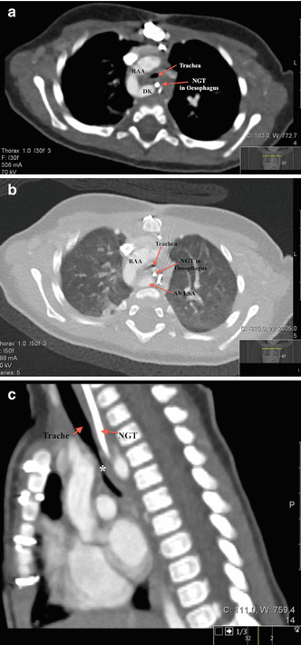
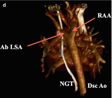
Fig. 19.2
(a) Axial image from the patient’s CT angiogram on a mediastinal window showing right aortic arch (RAA) with diverticulum of Kommerell (DK) and NGT inside the oesophagus. Significant compression of the trachea can be seen. (b) Axial image from the patient’s CT angiogram on a lung window showing the right aortic arch (RAA) and aberrant left subclavian artery (Ab LSA) compressing on the trachea and oesophagus. The trachea is severely narrowed. (c) Sagittal view of the patient’s CT angiogram showed that the trachea and oesophagus are compressed by the vascular ring causing severe narrowing (*). (d) 3D reconstruction of the CT angiogram from the posterior aspect to show the aberrant left subclavian artery (Ab LSA) and right aortic arch impinging on the oesophagus and trachea (oesophagus and trachea not shown; nasogastric tube (NGT) indicates position of oesophagus)
The patient underwent surgical repair of the vascular ring with transection and resection of Kommerell’s diverticulum and re-implantation of the left subclavian artery to the left common carotid artery. Recovery was uneventful and is now completely asymptomatic.
Vascular Ring
Background
Vascular ring is a congenital anomaly of the aortic arch and pulmonary artery that occurs early in development of the embryonic branchial arches. Due to their close spatial relationships with the trachea and oesophagus, abnormalities in the size, position and/or branching pattern of the aortic arch and pulmonary arteries can result in compression of these adjacent structures. This can lead to life-threatening respiratory distress signs, with or without oesophageal compression symptoms.
Dr Robert Gross first described a vascular ring in 1945 after he performed the first successful division of a double aortic arch. Incidence is only 1–3 % of all congenital heart disease. Given the risk of airway compromise, a high level of suspicion of these malformations must be maintained, as prompt diagnosis and treatment can be lifesaving.
Embryology
By the fourth week of embryogenesis, six symmetrical paired aortic arches and a pair of dorsal aortae are present in the embryo. Sequential fusion and regression of this complex structure results in the formation of a normal left aortic arch (i.e., an aortic arch which crosses over the left mainstem bronchus), descending on the left side of the spine. A normal left aortic arch gives rise to three branching vessels:
Innominate (brachiocephalic) artery, bifurcating into the right common carotid artery and the right subclavian artery
Left common carotid artery
Left subclavian artery
Persistence of the double arch results in the commonest form of vascular ring, which can be explained by the hypothetical double aortic arch model (Fig. 19.3a) proposed by Dr Jesse E. Edwards in 1948. According to his model, the ascending and descending aorta are connected by symmetrical arches on each side, forming a complete vascular ring around the trachea and oesophagus. Each aortic arch gives origin to common carotid and subclavian arteries. On each side, the corresponding ductus arteriosus connects the pulmonary artery and subclavian artery, forming an additional vascular ring.
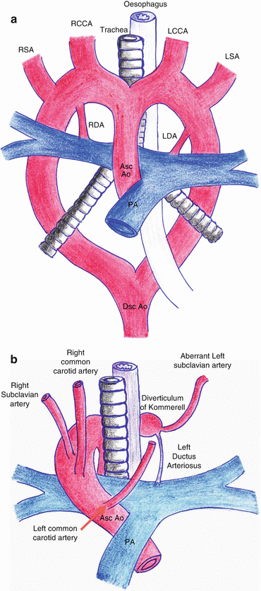


Fig. 19.3
(a) Schematic diagram of Edward’s hypothetical double aortic arch model. RSA right subclavian artery; RCCA right common carotid artery; LCCA left common carotid artery; LSA left subclavian artery; Asc Ao ascending aorta; Dsc Ao descending aorta; PA pulmonary artery. (b) Schematic diagram of a right aortic arch with aberrant left subclavian artery and left ductus arteriosus. The aberrant left subclavian artery can be seen arising from the diverticulum of Kommerell. Asc Ao ascending aorta; PA pulmonary artery
< div class='tao-gold-member'>
Only gold members can continue reading. Log In or Register to continue
Stay updated, free articles. Join our Telegram channel
Full access? Get Clinical Tree
