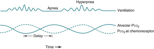
Disorders of Ventilatory Control

18


Disorders of Ventilatory Control


The finely tuned system of ventilatory control described in Chapter 17 is altered in a variety of clinical circumstances. In some cases, a primary disorder of the nervous system affects the neurologic network involved in ventilatory control and therefore may either diminish or increase the “drive” to breathe. In other instances the controlling system undergoes a process of adaptation in response to primary lung disease, so any alteration in function is a secondary phenomenon.


This chapter considers primary and secondary disturbances in ventilatory control. Of the secondary disorders, the most commonly seen is that associated with chronic obstructive pulmonary disease; therefore, the discussion of secondary disorders of ventilatory control focuses on this particular disorder. A common disturbance in the pattern of breathing, termed Cheyne-Stokes breathing, is covered, with a brief discussion of its pathogenesis. The final topic is ventilatory disorders associated with sleep, because alteration of ventilatory control may be an important component of the pathogenesis of sleep-related respiratory dysfunction.


Primary Neurologic Disease


Several diseases of the nervous system alter ventilation, apparently by affecting regions involved in ventilatory control. However, the results are variable, depending on the type of disorder and the region involved. In some cases hyperventilation is prominent, whereas in others hypoventilation is significant. In a third category the most apparent change occurs in the pattern of breathing.


Presentation with Hyperventilation


With certain acute disorders of the central nervous system (CNS), hyperventilation (i.e., decreased PCO2 and respiratory alkalosis) is relatively common. Acute infections (meningitis, encephalitis), strokes, and trauma affecting the CNS are notable examples. The exact mechanism of hyperventilation in these situations is not known with certainty. Patients with hyperthyroidism frequently present with hyperventilation that resolves after treatment. Increased sensitivity of the chemoreceptors in the brain during hyperthyroidism appears to account for the effect. Hyperventilation frequently complicates advanced hepatic disease, presumably because of increased concentrations of circulating substances stimulating ventilation that normally are metabolized by the healthy liver. Some proposed substances causing central stimulation of respiration in patients with hepatic disease include progesterone, ammonia, and glutamate.


Presentation with Hypoventilation


A presentation with hypoventilation presumably results from a primary insult to the nervous system that affects centers involved with control of breathing. In such circumstances, patients have an elevated PCO2, but because the clinical problems are generally not acute, the pH level has returned closer toward normal as a result of renal compensation with retention of bicarbonate. When no specific etiologic factor or prior event can be found to explain the hypoventilation, the patient is said to have idiopathic hypoventilation or primary alveolar hypoventilation. Other patients have suffered a significant insult to the nervous system at some time in the past (e.g., encephalitis), and chronic hypoventilation presumably is a sequela of the past event.


Patients with these syndromes of hypoventilation are characterized by depressed ventilatory responses to the chemical stimuli of hypercapnia and hypoxia. Measurement of arterial blood gases generally reveals an elevation in arterial PCO2 accompanied by a decrease in PO2, the latter primarily attributable to hypoventilation. As in other disorders associated with these blood gas abnormalities, cor pulmonale may result and be the presenting problem in these syndromes. The term congenital central hypoventilation syndrome or Ondine’s curse (see Chapter 17) has been applied to a rare subset of patients with congenital alveolar hypoventilation. However, an element of decreased ventilatory response to hypercapnia and hypoxia is much more commonly seen in clinical practice and probably represents a spectrum of abnormalities in ventilatory response.


In the past, treatment of alveolar hypoventilation generally centered around two modalities: drugs (most commonly the hormone progesterone) and electrical stimulation of the phrenic nerve. Progesterone is well known to be a respiratory stimulant and in some cases may improve respiratory drive and decrease CO2 retention. In the second approach, the diaphragm can be induced to contract by repetitive electrical stimulation of the phrenic nerve, which can be achieved by intermittent current applied via an implanted electrode. Although both of these modalities are still used, the most common current therapy for patients with clinically significant hypoventilation is noninvasive positive-pressure (i.e., assisted) ventilation, usually applied nocturnally. This topic is discussed in Chapter 29.


Cheyne-Stokes Breathing


Cheyne-Stokes breathing is a cyclic pattern in which periods of gradually increasing ventilation alternate with periods of gradually decreasing ventilation (even to the point of apnea). This type of ventilation is shown schematically in Figure 18-1. It has been known for many years that two main types of disorders are associated with this type of breathing: heart failure and some forms of CNS disease. Cheyne-Stokes breathing can also be seen under certain physiologic situations even in the absence of underlying disease. Examples include the onset of sleep and exposure to high altitude.


Central to the pathogenesis of Cheyne-Stokes ventilation is a problem with the feedback system of ventilatory control. Normally the controlling system can adjust its output to compensate for arterial blood gas values that differ from the ideal or desired state. For example, with an elevated arterial PCO2, the central chemoreceptor signals the medullary respiratory center to increase its output to augment ventilation and restore PCO2 to normal. Similarly, the peripheral chemoreceptor responds to hypoxemia by increasing its output, signaling the medullary respiratory center to augment ventilation and restore PO2 to normal.


At times, this feedback system may fail, especially if there is a delayed response to the signal or if the system responds more than necessary and overshoots the mark. Such defects in the feedback process appear to be at work in Cheyne-Stokes breathing. This section touches on a few aspects of theories proposed to explain Cheyne-Stokes ventilation. For further discussion, the interested reader is referred to the references.


Prolongation in circulation time, which is one mechanism postulated to play a role in heart failure, results in an abnormal delay between events in the lung and sensing of PCO2 changes by the central chemoreceptors. Hence, medullary respiratory output is out of phase with gas exchange at the lungs, and oscillations in ventilation occur as the central chemoreceptor and the medullary respiratory center make belated attempts to maintain a stable PCO2 (see Fig. 18-1).


An alternative explanation for Cheyne-Stokes breathing that occurs with heart failure is an accentuated ventilatory response to hypercapnia. This type of heightened responsiveness of the feedback system produces “instability” of respiratory control and a cyclic overshooting and undershooting of ventilation. Such increased responsiveness of the ventilatory control system may also play a role in patients with CNS disease who exhibit periods of Cheyne-Stokes respiration.


A similar type of instability of ventilatory control occurs when hypoxia is driving the feedback system, as is seen on exposure to high altitude. The ventilatory response to hypoxia is nonlinear. For the same drop in PO2, the increment in ventilation is larger at a lower absolute PO2 (see Fig. 17-3). This means that at a relatively high initial PO2, the system is less likely to respond to small changes in PO2 but then is apt to overshoot as PO2 falls further. This instability of the respiratory control system results in a widely oscillating output from the respiratory center and thus a cyclic pattern of ventilation.


Control Abnormalities Secondary to Lung Disease


Ventilatory control mechanisms often respond to various forms of primary lung disease by altering respiratory center output. Either stimulation of peripheral chemoreceptors by hypoxemia or stimulation of receptors by diseases affecting the airways or pulmonary interstitium can induce the respiratory center to increase its output, resulting in respiratory alkalosis. For example, patients with asthma commonly demonstrate increased respiratory drive and hyperventilation during acute attacks as a consequence of stimulation of airway receptors. Similarly, patients with acute pulmonary embolism, pneumonia, or chronic interstitial lung disease often hyperventilate, presumably as a result of stimulation of one or more types of intrathoracic receptors, with or without the additional ventilatory stimulus contributed by hypoxemia.


In contrast, patients with chronic obstructive pulmonary disease (COPD) have variable levels of PCO2. Some patients with COPD do not demonstrate CO2

Stay updated, free articles. Join our Telegram channel

Jun 12, 2016 | Posted by in RESPIRATORY | Comments Off on Disorders of Ventilatory Control

Full access? Get Clinical Tree

Get Clinical Tree app for offline access