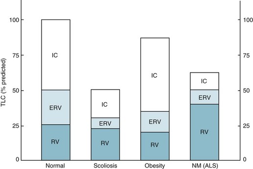
Disorders of the Respiratory Pump

19


Disorders of the Respiratory Pump


The chest wall, diaphragm, and related neuromuscular apparatus moving the chest wall act in concert to translate signals from the ventilatory controller into expansion of the thorax. Together these structures constitute the respiratory pump, an important system that may fail as a result of diseases affecting any of its parts. Because disorders of the respiratory pump include a variety of problems, this discussion is limited to those disorders that are most common and most important clinically: (1) neuromuscular disease affecting the muscles of respiration (Guillain-Barré syndrome, myasthenia gravis, poliomyelitis, and amyotrophic lateral sclerosis), (2) diaphragmatic fatigue, (3) diaphragmatic paralysis, and (4) diseases affecting the chest wall (kyphoscoliosis, obesity).


Neuromuscular Disease Affecting the Muscles of Respiration


A number of neuromuscular diseases have the potential for affecting the muscles of respiration. In some cases, the underlying process is acute and generally reversible (e.g., Guillain-Barré syndrome), and the muscles of respiration are transiently affected for a variable amount of time. In other cases, the neuromuscular damage is permanent, and any consequences that impair function of the muscles of respiration are chronic and irreversible. This chapter provides brief definitions of some specific neurologic disorders with respiratory sequelae, followed by a discussion of the pathophysiology and clinical consequences of these diseases as they relate to the respiratory system.


Specific Diseases


The major neuromuscular diseases that can affect the muscles of respiration are listed in Table 19-1; several are discussed here.


Guillain-Barré syndrome is a disorder characterized by demyelination of peripheral nerves. It is thought to be triggered by exposure to an antigen (typically an infectious agent such as Campylobacter jejuni). The resulting immune response is misdirected to similar antigenic determinants (epitopes) on neural tissue or Schwann cells. Patients frequently have a history of a recent viral or bacterial illness followed by development of an ascending paralysis and variable sensory symptoms. Classically, weakness or paralysis starts symmetrically in the lower extremities and progresses or ascends proximally to the upper extremities and trunk. In up to one-third of cases, the disease is more severe, with respiratory muscle weakness or paralysis accompanying the more usual limb and trunk symptoms. When respiratory muscles are affected, respiratory failure often supervenes but usually is reversible over the course of weeks to months. Generally, the natural history of the disease leads to recovery, although 3% to 8% of patients die, and up to 10% of survivors have permanent sequelae.


In myasthenia gravis, patients experience weakness and fatigue of voluntary muscles, most frequently those innervated by cranial nerves, but peripheral (limb) and, potentially, respiratory muscles also are affected. The primary abnormality is found at the neuromuscular junction, where transmission of impulses from nerve to muscle is impaired by a decreased number of receptors on the muscle for the neurotransmitter acetylcholine and by the presence of antibodies against these receptors. Although myasthenia gravis is a chronic illness, the manifestations often can be controlled by appropriate therapy, and individual episodes of respiratory failure are potentially reversible.


Poliomyelitis is a viral disease in which the poliovirus attacks motor nerve cells of the spinal cord and brainstem. Both the diaphragm and intercostal muscles can be affected, with resulting weakness or paralysis and respiratory failure. Surviving patients generally recover respiratory muscle function, although some patients have chronic respiratory insufficiency from prior disease. Mass vaccination of the population in developed countries makes new cases rare. In postpolio syndrome, patients develop new or progressive symptoms of weakness that occur decades after the initial episode of poliomyelitis. Involvement occurs in muscles originally affected by the disease, so respiratory muscle involvement is more likely in patients who had respiratory failure with their initial disease.


Amyotrophic lateral sclerosis is a degenerative disease of the nervous system that involves both upper and lower motor neurons. Commonly, muscles innervated by either cranial nerves or spinal nerves are affected. Clinically, progressive muscle weakness and wasting develop, eventually leading to profound weakness of respiratory muscles and death. Although the time course of the disease is variable among patients, the natural history is one of irreversibility and progressive deterioration. As a result, patients and families must confront the difficult decision of whether to use mechanical ventilation either noninvasively or through a tracheostomy tube when the patient is in respiratory failure, knowing that no treatment will arrest the progressive neurologic deterioration.


Pathophysiology and Clinical Consequences


Weakness of respiratory muscles is the hallmark of respiratory involvement in the neuromuscular diseases. Depending on the specific disease, chest wall (intercostal) muscles, diaphragm, and expiratory muscles of the abdominal wall are affected to variable extents.


Impairment of inspiratory muscle strength may render patients unable to maintain sufficient minute ventilation for adequate CO2 elimination. In addition, patients often alter their pattern of breathing, taking shallower and more frequent breaths. Although this pattern of breathing may require less muscular effort and be more comfortable, it is also less efficient because a greater proportion of each breath is wasted on ventilating the anatomic dead space (see Chapter 1). Therefore, even if total minute ventilation is maintained, alveolar ventilation (and thus CO2 elimination) is impaired by the altered pattern of breathing.


The respiratory difficulty that develops in patients with neuromuscular disease is complicated by weakness of expiratory muscles and by an ineffective cough. Recurrent respiratory tract infections, accumulation of secretions, and areas of collapse or atelectasis contribute to the clinical problems seen in these patients.


Symptoms include dyspnea and anxiety. Patients may also have a feeling of suffocation. Often the presence of generalized muscle weakness severely limits patients’ activity and lessens the degree of dyspnea that would be present if they were capable of more exertion.


With severe neuromuscular disease, pulmonary function tests show a restrictive pattern of impairment. Although muscle weakness is the primary cause of restriction, compliance of the lung and chest wall may be secondarily affected, further contributing to the restrictive pattern. The decrease in pulmonary compliance presumably is due to microatelectasis (i.e., multiple areas of alveolar collapse) resulting from the shallow tidal volumes. At the same time, stiffening of various components of the chest wall (e.g., tendons, ligaments, and joints) over time is thought to be responsible for decreased distensibility of the chest wall. Functional residual capacity (FRC) is normal or decreased, depending on how much respiratory system compliance is altered. Total lung capacity is decreased primarily as a result of inspiratory muscle weakness, but changes in respiratory system compliance may contribute as well. Residual volume (RV) frequently is increased as a result of expiratory muscle weakness (Fig. 19-1). The degree of muscle weakness can be quantified by measuring the maximal inspiratory and expiratory pressures the patient is able to generate with maximal inspiratory and expiratory efforts against a closed mouthpiece. Both maximal inspiratory pressure (MIP) and maximal expiratory pressure may be significantly depressed.


In the setting of severe muscle weakness, arterial blood gases are most notable for the presence of alveolar hypoventilation (i.e., hypercapnia). Hypoxemia due to alveolar hypoventilation and the associated depression in alveolar PO2 also occurs. When hypoventilation is the sole cause of hypoxemia, the alveolar-arterial oxygen difference (AaDO2) is normal. However, complications such as atelectasis, respiratory tract infections, and inadequately cleared secretions may add a component of ventilation-perfusion mismatch or shunt that further depresses PO2 and increases AaDO2.


Diaphragmatic Disease


Although diaphragmatic involvement is a significant component of many of the neuromuscular diseases that affect the muscles of respiration, additional etiologic and clinical considerations justify a separate discussion of diaphragmatic disease. First we consider diaphragmatic fatigue, a potential consequence of disorders affecting other parts of the respiratory system that significantly increase the workload placed on the diaphragm. We then discuss diaphragmatic paralysis, with separate considerations of unilateral and bilateral paralysis, because the causes and clinical manifestations are often quite different.


Diaphragmatic Fatigue


Excluding cardiac muscle, the diaphragm is the single muscle used most consistently and repetitively throughout the course of a person’s lifetime. It is well suited for sustained activity and aerobic metabolism, and under normal circumstances the diaphragm does not become fatigued.


However, if the diaphragm is required to perform an excessive amount of work or if its energy supplies are limited, fatigue may develop and may contribute to respiratory dysfunction in certain clinical settings. For example, if a healthy individual repetitively uses the diaphragm to generate 40% or more of its maximal force, fatigue develops and prevents this degree of effort from being sustained indefinitely. For patients with diseases that increase the work of breathing, particularly obstructive lung disease and diseases of the chest wall (described in the section on diseases affecting the chest wall), the diaphragm works at a level much closer to the point of fatigue. When a superimposed acute illness further increases the work of breathing or when an intercurrent problem (e.g., depressed cardiac output, anemia, or hypoxemia) decreases the energy supply available to the diaphragm, diaphragmatic fatigue may contribute to the development of hypoventilation and respiratory failure.


Inefficient diaphragmatic contraction is another factor that may contribute to diaphragmatic fatigue, especially in patients with obstructive lung disease. When the diaphragm is flattened and its fibers are shortened as a result of hyperinflated lungs, the force or pressure developed during contraction is less for any given level of diaphragmatic excitation (see Chapter 17). Therefore, a higher degree of stimulation is necessary to generate comparable pressure by the diaphragm, and increased energy consumption results.

Only gold members can continue reading. Log In or Register to continue

Stay updated, free articles. Join our Telegram channel

Jun 12, 2016 | Posted by in RESPIRATORY | Comments Off on Disorders of the Respiratory Pump

Full access? Get Clinical Tree

Get Clinical Tree app for offline access