Chapter 64 Diseases of the Thoracic Cage and Respiratory Muscles
Dyspnea and alveolar hypoventilation occur in patients with restrictive lung disease as a consequence of an imbalance between the respiratory muscle load and respiratory muscle capacity. Subsequent modification of the neural respiratory drive occurs, which directly reflects this imbalance. This chapter begins with an overview of the anatomy of the respiratory muscle pump and includes a detailed explanation of the pathophysiology of chronic respiratory failure. It provides a comprehensive guide to the physician of how best to assess and investigate a patient with suspected respiratory compromise from respiratory muscle weakness, chest wall disease, or obesity. The second part of the chapter focuses on specific neurologic/neuromuscular and chest wall diseases associated with respiratory impairment.
Respiratory Muscle Pump
The respiratory system is made up of two main components: the respiratory muscle pump, which facilitates airflow, thereby enabling ventilation, and the lungs, which support pulmonary gas exchange (see Chapter 6 for more detailed explanations and for respiratory muscle testing). The respiratory muscle pump itself is made up of the inspiratory muscles, the diaphragm and extradiaphragmatic accessory muscles, and the expiratory muscles, principally the abdominal wall muscles. Inspiration is an active process in which contraction of the diaphragm forces the abdominal contents in an anterocaudal direction to allow the volume of the chest cavity to increase. This expansion, in turn, generates a negative subatmospheric pressure to form a pressure gradient that drives air into the lungs. At rest, expiration is a passive process with the elastic properties of the chest wall and lungs providing the recoil forces that allow the lung volume to return to the functional residual capacity. During exercise and forced expiratory maneuvers (e.g., coughing), however, contraction of the abdominal muscles occurs, pushing the diaphragm upward in the absence of flow limitation.
Clinical Consequences of Respiratory Muscle Pump Imbalance
Breathlessness, alveolar hypoventilation and hypercapnic respiratory failure result from an imbalance between the respiratory muscle load, capacity, and neural respiratory drive (Figure 64-1). By using this model the physician can develop a simple clinical tool to assess the breathless patient and the patient in chronic respiratory failure. More commonly, in patients with restrictive lung disease, the reduction in capacity occurs as a consequence of intrinsic muscle weakness. On occasion, however, capacity may be relatively preserved, but in the context of a significant respiratory muscle load or poor orientation of the inspiratory muscles (e.g., kyphoscoliosis), this may limit the pressure-generating capacity, so that dyspnea and alveolar hypoventilation can develop subsequently. In critically ill patients, the respiratory muscle weakness may be transient and reversible, such as occurs in respiratory acidosis, hypokalemia, and hypophosphatemia (Box 64-1).
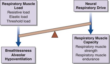
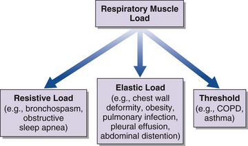
An increase in respiratory muscle load occurs in patients with increased airway resistance, chest wall deformity, or obesity. Any of these conditions will accelerate the onset of dyspnea and alveolar hypoventilation in patients with respiratory muscle weakness (Figure 64-2).
In healthy subjects, a significant reduction in neural respiratory drive to skeletal muscle, including the respiratory muscles, occurs during sleep. Despite the complete loss of skeletal muscle activity during rapid eye movement (REM) sleep, in normal subjects significant nocturnal hypoxia and hypercapnia are avoided because the activation of the diaphragm to maintain ventilation is preserved. By contrast, in patients with respiratory muscle weakness, especially diaphragmatic weakness and paralysis, hypoventilation during REM sleep often is the first sign of declining respiratory function. Of interest, in patients with idiopathic diaphragmatic paralysis, hypoventilation is prevented by a neuroadaptive mechanism with cyclic activation of the sternocleidomastoid muscle during REM sleep, which maintains ventilation. Also, there is substantial reserve in the respiratory muscle pump such that inspiratory muscle strength must fall to one third of normal before the onset of respiratory failure. Furthermore, clinical deterioration is observed at an earlier stage if an increased load is applied to the system, such as in pneumonia, or if the neural respiratory drive is modified with drugs such as benzodiazepines, opiates, and other anesthetic agents used during routine anesthesia.
Assessment of the Respiratory Muscle Pump
The respiratory muscle pump is essential for effective ventilation and to maintain gas exchange. In addition to a directed clinical history and examination, the physician can use simple bedside tests as well as more advanced measurements of respiratory muscle function to perform a detailed assessment of the patient with restrictive respiratory muscle and chest wall disease.
Clinical Features
As always, a detailed history will facilitate making a correct diagnosis (Table 64-1). Of particular importance is the rate of onset of symptoms. Acute respiratory muscle weakness tends to progress rapidly, culminating in a medical emergency requiring intubation and mechanical ventilation. Some causes of acute respiratory muscle weakness, such as an acute myasthenic crisis or Guillain-Barré disease, may be reversible, and it is important to facilitate invasive respiratory support in the acute setting until a diagnosis can be made and appropriate therapies initiated. In chronic respiratory failure, the onset may not be clear, but clues to the course of events can be gained from the history. Often patients will describe dyspnea on exertion, but if peripheral muscle weakness precedes respiratory muscle weakness, the consequent loss of significant locomotor function will not permit exertion of sufficient degree to produce dyspnea. Classic features of diaphragm weakness are not always present in patients with generalized neuromuscular disease but may include dyspnea on lying supine, on bending forward, or on immersion in water above the midchest level. If respiratory weakness is severe, patients also may describe symptoms of nocturnal hypoventilation, such as extensive daytime somnolence, reduced concentration, and morning headaches that typically resolve within 30 minutes of waking. Specifically, in the pediatric and adolescent populations, clinicians should be alerted to more subtle clinical features such as failing school performance, recurrent episodes of chest sepsis, reduction in appetite, and weight loss.
Table 64-1 Important Clinical Features in Diseases of the Thoracic Cage and Respiratory Muscles
Disorder | Clinical Manifestations |
---|---|
Sleep-disordered breathing | Morning headache, daytime sleepiness, disrupted sleep pattern, impaired intellectual function, generalized fatigue, loss of appetite and weight |
Respiratory muscle weakness | Orthopnea, breathlessness on immersion in water, breathlessness on leaning forward, breathlessness on exertion, poor cough, poor chest expansion, paradoxical abdominal motion during inspiration (inward motion of the anterior abdominal wall due to diaphragm weakness), abdominal muscle recruitment in expiration |
Bulbar dysfunction | Low volume voice, difficulty swallowing, drooling, difficulty clearing secretions, poor cough, staccato/slurred speech, coughing on swallowing |
In all cases of respiratory muscle weakness, an important aspect of the evaluation is to identify any symptoms that may suggest generalized neuromuscular weakness—in particular, decrements in the patient’s speech and swallowing function, as well as weakness of the arms and legs. Weakness of the abdominal muscles may result in difficulty achieving an effective cough to clear secretions and debris from the airways. This impairment leads to issues with sputum retention and increases the risk of chest infection. In the case of chest wall disease, the common causes kyphoscoliosis and obesity usually are self-evident. As highlighted earlier, certain drugs have effects that can accelerate respiratory decline, so attention should be given to the common offenders, such as benzodiazepines, opiates, and corticosteroids. More recently, in particular, in the Duchenne muscular dystrophy (DMD) population, corticosteroids are increasingly being used to maintain locomotor muscle function, and these agents are associated with substantial weight gain and consequent upper airway obstruction. Clinicians must be aware of this potential problem in this younger patient population.
Physical examination requires a careful and considered approach, which must include a thorough neurologic examination to observe for signs of tongue and peripheral muscle fasciculation, peripheral muscle wasting and weakness, and peripheral sensory loss. Other signs, such as pseudohypertrophy of the calf muscles, are observed in patients with muscular dystrophies. Scars of previous operations may indicate possible trauma to underlying neuromuscular structures or an imposed restrictive chest wall deformity, such as from a phrenic nerve crush, thoracoplasty, coronary artery bypass grafting, thyroidectomy, or thymoma resection. With severe diaphragm weakness, abdominal inspiratory paradox is observed: The anterior abdominal wall moves inward as a result of the failure of a weak or paralyzed diaphragm to descend against the force exerted by the abdominal contents. With more generalized weakness, global loss of thoracic expansion on inspiration is observed.
Arterial Blood Gas Analysis
Although arterial blood gas analysis is one of the most common first-line investigations to diagnose chronic respiratory failure, limitations associated with this technique have been recognized. In particular, the daytime partial pressure of carbon dioxide (PaCO2) may be normal despite the presence of substantial inspiratory muscle weakness, nocturnal hypoventilation, and nocturnal hypercapnia. Measurement of arterial blood gas partial pressures is essential, however, to define the severity of respiratory failure. Most commonly, hypoventilation caused by neuromuscular respiratory weakness and chest wall disease manifests with a mild hypoxia, with a partial pressure of oxygen (PaO2) less than 60 mm Hg, and hypercapnia, with a PaCO2 greater than 45 mm Hg.
The respiratory failure associated with many neurologic and chest wall conditions frequently manifests insidiously, with hypercapnia triggering metabolic compensation and renal retention of bicarbonate to maintain the hydrogen ions and, consequently, arterial pH within the normal range.
In the event of rapid onset of neurologic symptoms, such as in Guillain-Barré syndrome and myasthenic syndrome, or with acute deterioration of chronic respiratory failure, there is less time for renal buffering, so hypercapnic respiratory acidosis will occur.
Pulmonary Function Tests
Patients with respiratory neuromuscular weakness and those with chest wall disease both are characterized by a reduced ability to expand the thoracic rib cage. In turn, this failure to expand the rib cage results in a lack of generation of sufficient negative intrathoracic pressure to facilitate adequate inspiratory airflow. As a result, these patients present with clinical features of a restrictive lung function pattern characterized by a reduced forced vital capacity (FVC) and an elevated ratio of forced expiratory volume in 1 second (FEV1) to FVC (FEV1/FVC greater than 80%). Although vital capacity (VC) is in widespread use and is relatively simple to determine, its utility is limited because it has low sensitivity. In particular, significant inspiratory muscle weakness is required before a significant fall in VC is observed. A fall in VC on adoption of the supine position is specific for respiratory muscle weakness, and a fall in VC of more than 20% suggests bilateral diaphragmatic weakness. Of importance, the VC can be preserved in persons with a moderate degree of global respiratory muscle weakness or hemidiaphragm weakness.
The pattern of respiratory muscle weakness must be considered in interpreting results of pulmonary function tests in patients with chest wall disease and respiratory muscle weakness. Although total lung capacity (TLC) and VC are reduced in patients with predominantly inspiratory muscle weakness and combined inspiratory and expiratory muscle weakness, the pattern of weakness influences functional residual capacity (FRC), residual volume (RV), overall gas transfer (DLCO), and gas transfer corrected for alveolar volume (Table 64-2).
Table 64-2 Pulmonary Function Tests for Patients with Predominantly Inspiratory Muscle Weakness and Combined Inspiratory/Expiratory Muscle Weakness
Inspiratory Muscle Weakness | Combined Inspiratory/Expiratory Muscle Weakness |
---|---|
Reduced VC | Reduced VC |
Fall in supine VC >20% | N/A |
FEV1/FVC ratio >80% | FEV1/FVC ratio >80% |
Reduced TLC | Reduced TLC |
Normal RV | Increased RV |
Reduced FRC | Increased FRC |
Reduced TLCO | Reduced TLCO |
“Supranormal” KCO | Reduced KCO |
FEV1, forced expiratory volume in 1 second FRC, functional residual capacity; FVC, forced vital capacity; KCO, gas transfer coefficient corrected for alveolar volume; N/A, not available; RV, residual volume; TLC, total lung capacity; TLCO, overall gas transfer; VC, vital capacity.
Tests of Respiratory Muscle Function
Because respiratory muscle contraction generates tension, the consequent pressure change that occurs can serve as an in vivo measure for the quantification of respiratory muscle strength. In addition, recent developments have expanded the current understanding of the mechanical actions and interactions of the respiratory muscles. A greater emphasis has been placed on assessing and quantifying respiratory muscle strength and pulmonary mechanics in a variety of patient groups, including children, adolescents, and adults (see Chapter 6 for a more detailed discussion).
Maximal Inspiratory and Expiratory Pressures
Maximal inspiratory and expiratory pressure measurements are clinically useful noninvasive tests with established reference ranges. As with any volitional test, however, results will be dependent on subject motivation and maximal effort, which explains in part the wide range of normal values. Furthermore, the observed pressure depends on mouthpiece design and patient posture, and it often is difficult to distinguish between mild weakness and normal strength on an individual basis. Nevertheless, maximum inspiratory mouth pressure (PImax) can provide a simple rapid estimation of global inspiratory muscle strength (in men, below −80 cm H2O; in women, below −70 cm H2O), but it does not allow specific conclusions to be drawn about the function of the diaphragm. If the maneuver is performed from FRC, PImax reflects the strength of the inspiratory muscles, whereas with performance from RV, the test will be influenced by the elastic recoil of the chest wall. In clinical practice, patients find it easier to perform the test from FRC rather than RV, and previous data have shown little difference between the peak or plateau values measured from either RV or FRC.
The strength of the expiratory muscles, principally the abdominal muscles, is assessed by measuring the static expiratory pressure generated at the mouth (PEmax). As with the PImax, the range of the normal values is wide (in men, above +130 cm H2O; in women, above +100 cm H2O) and some patients can find this maneuver difficult to perform, particularly those patients with weakness of the orofacial muscles. Thus, as with PImax, although a high value of PEmax excludes expiratory muscle weakness, a low value can be difficult to interpret. PEmax are can be measured from either TLC or FRC. PImax and PEmax measurements are reduced in females and decline with age.
Sniff Inspiratory Pressure
The rapid inspiratory effort of a sniff maneuver (see Chapter 6) is accompanied by momentary equilibration of intrathoracic and upper airway pressures. This equilibration occurs above a pressure of 10 to 12 cm H2O, so employing sniff nasal pressure (Pnsn) allows noninvasive measurement of inspiratory muscle strength. Pnsn is a particularly useful additional investigation in patients with a low or equivocal PImax to confirm or exclude the presence of inspiratory muscle weakness.
Cough Gastric Pressure
As with sniffing, patients find coughing a natural maneuver. Although assessment of the cough gastric pressure (Pgascough) requires the insertion of a gastric balloon, the measurement is easy to learn and more reproducible than with PEmax. Although normal values for Pgascough have been reported, previous studies using Pgascough as a marker for expiratory muscle strength are described in the literature. Normal Pgascough values are above +215 cm H2O in men and above +165 cm H2O in women.
Cough Peak Flow
Cough peak flow is a simple noninvasive measure of expiratory muscle function, which is considered to be a useful marker of cough function and a reflection of the patient’s ability to clear respiratory secretions. Values of less than 160 L/min are reported to be the cutoff level for increased risk of developing chest sepsis. A cough peak flow less than 160 L/min can be augmented by the mechanical insufflator-exsufflator device used with a face mask.
Maximum Transdiaphragmatic Pressure Measurements
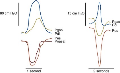
The previously described tests provide data only on global respiratory muscle function, rather than on diaphragm function itself. To specifically assess diaphragm function, transdiaphragmatic pressure (Pdi) is measured by placement of a pressure-monitoring catheter in both the esophagus and stomach, to provide measurement of the esophageal and gastric pressures (Figure 64-3).
Although transdiaphragmatic pressure during a maximal static maneuver (Pdimax) has been reported, this maneuver gives a wide normal range in naive (i.e., previously untested) subjects. An alternative is to obtain maximal sniff transdiaphragmatic pressure (Pdisn), which has gained recognition in clinical practice not only because it is a familiar maneuver but because it measures both Pessn and Pdisn, providing data on global inspiratory muscle and diaphragm strength (Figure 64-4).

Full access? Get Clinical Tree

