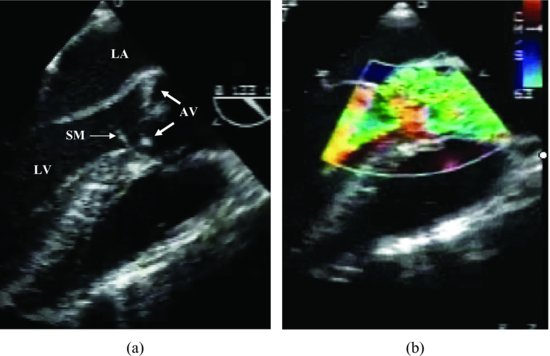
TEE images indicate a subaortic membrane, which can be well seen during diastole and systole with turbulent flow in left ventricular outflow tract (LVOT) (Figure 51.2, Videoclip 51.1).
Figure 51.2 Transesophageal echocardiogram images indicate a subaortic membrane (arrow) (a), which can be well seen with aortic valve stenosis turbulent flow in left ventricular outflow tract during systole (b). AV, aortic valve; LA, left atrium; LV, left ventricle; SM, subaortic membrane.

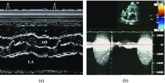
M-mode recording from our case with discrete subaortic stenosis shows the typical early systolic notch followed by high-frequency fluttering of the valve leaflet throughout the remainder of ejection period. Continuous wave Doppler recording indicates high-velocity flow through LVOT (Figure 51.3).
Figure 51.3 M-mode recording from our case with discrete subaortic stenosis shows the typical early systolic notch (arrow) followed by high frequency fluttering of the valve leaflet throughout the remainder of ejection period (a). Continuous wave Doppler recording indicates high-velocity flow through left ventricular outflow tract (b). AO, aorta; LA, left atrium.

The right ventricular size and function are normal.
Stay updated, free articles. Join our Telegram channel

Full access? Get Clinical Tree


