21 Diffuse Lung Diseases
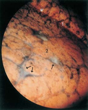
Fig. 21.1
Sarcoidosis stage II. After induction of a right-sided pneumothorax, in the interlobular septae, anthracotic pigment is seen outlining subpleural lymphatics. Upper lobe (1). In the middle lobe (2) there is whitish thickening of the interstitium, in part stellate and in part geographic (→).
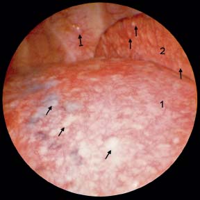
Fig. 21.2
Sarcoidosis stage II with pleural lesion. After induction of a left-sided pneumothorax, the lung (1) shows nodular lesions (→) on the surface of the left lower lobe (1) and upper lobe (2); on the chest wall is one typical sarcoid nodule ( ).
).
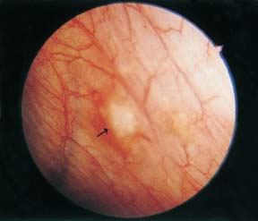
Fig. 21.3
Sarcoidosis stage II with pleural lesions. After induction of a left pneumothorax, the parietal pleura shows increased numbers of dilated vessels. In some places (→), whitish nodules, surrounded by a hyperemic zone protrude into the pleural space.
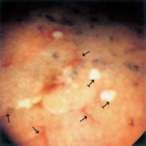
Fig. 21.4
Sarcoidosis stage III. After induction of a right pneumothorax, the interlobar regions are hyperemic (→). In all lobes there are small whitish nodules of 2-3 mm diameter without pigmented edge ( ).
).
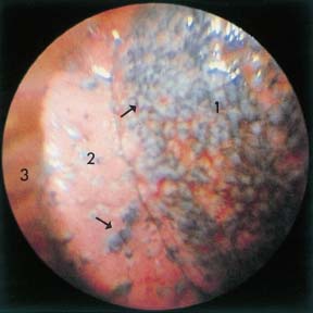
Fig. 21.5
Sarcoidosis stage III. After induction of a right pneumothorax, the lung is seeded with whitish nodules containing anthracotic pigment (→). Right upper lobe (1), interlobar pleura of the lower lobe (2), chest wall and ribs (3).
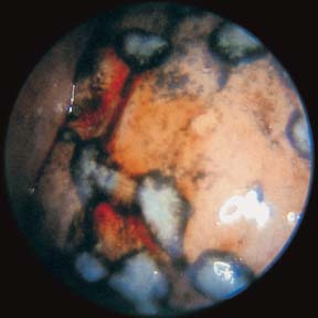
Fig. 21.6
Sarcoidosis stage III. After induction of a right pneumothorax, scattered over all lung lobes are lentil- to pea-sized, firm, whitish nodules containing bluish pigment.
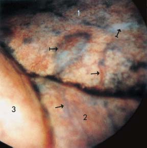
Fig. 21.7
Langerhans cell histiocytosis. After induction of a right pneumothorax, all lobes show distinct thickening of the interlobar septae (→) interspersed with small pale blebs ( ) covered with vessels. Upper lobe (1), middle lobe (2), lower lobe (3). Interlobar fissures clear.
Stay updated, free articles. Join our Telegram channel

Full access? Get Clinical Tree


