The incidence of end-stage renal disease (ESRD) has increased owing to the aging of our population and the increasing prevalence of diabetes. ESRD was a fatal illness until the establishment of chronic hemodialysis in the 1960s. Since then, the life expectancy of patients with ESRD has increased dramatically. As a result, there are nearly half a million ESRD patients in the United States today, the majority of whom are maintained by chronic hemodialysis (the remainder undergo peritoneal dialysis).1 The lives of hemodialysis patients depend on functional access to their circulation that provides an adequate volume of blood flow for the dialysis process, approximately 600 mL/min. Creation and maintenance of dialysis access is the leading cause of hospitalizations of these patients and costs the health care system in excess of a billion dollars annually. These costs can be reduced by increased use of native veins for dialysis access fistulas and better approaches to maintenance of patency—areas in which duplex scanning can play a major role. Duplex ultrasound helps identify veins for fistula creation that may otherwise be overlooked, ensures adequacy of arterial inflow and venous outflow, and aids in determining adequacy of fistula maturation and identification of reasons for maturation failure. Duplex scanning can also diagnose problems related to maintenance of fistulas, including arterial steal, pseudoaneurysms, infections, and stenoses in the access or venous outflow. This chapter reviews dialysis access and the role that duplex scanning plays in this process.
TYPES OF DIALYSIS ACCESS
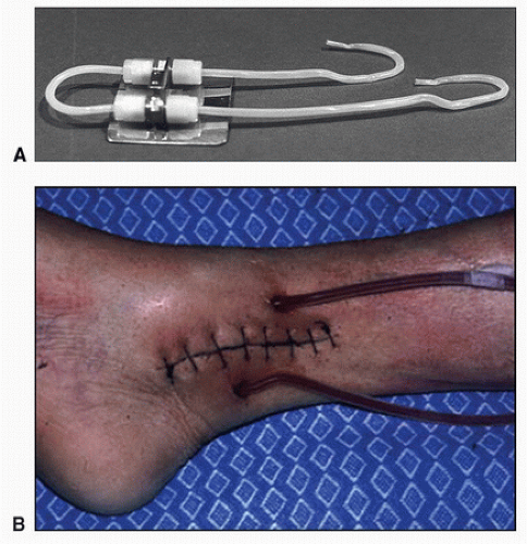
Maintenance hemodialysis was made possible by development of the Scribner shunt in 1960 by Scribner, Dillard, and Quinton in Seattle.2 These expanded polytetrafluoroethylene (ePTFE) shunts were placed percutaneously into a peripheral artery and vein (typically at the wrist or ankle) with tubing exiting the skin (Fig. 33.1). This allowed direct connection of arterial flow into the dialysis machine and venous return back to the patient’s circulation. When not in use, the two ePTFE tubes were connected to each other, creating a loop fistula outside the body. These devices were not suitable for long-term access because they were awkward, limited use of the extremity, and were plagued by frequent thrombosis and infection. The most common use of these devices was to support patients while recovering from acute renal failure. The later development of native arteriovenous fistulas by Cimino and Brescia,3 who described surgical creation of a fistula between the radial artery and the cephalic vein in 1962, provided the first durable method that could support chronic dialysis access. When arterialized, the cephalic vein dilates and thickens, allowing high-flow access to the bloodstream by direct needle puncture of this vein. This process of maturation takes about 8 weeks or longer, although some centers have advocated earlier use in the case of larger veins. To this day, the Cimino-Brescia fistula remains the first choice for access owing to its durability, low morbidity, and freedom from infection. In the years since the original description of the radiocephalic fistula, surgeons have found that nearly any adequately sized upper extremity vein can be connected to an artery to create a similar fistula, and deep veins can be transposed to a superficial location to allow percutaneous access. Most patients, even those whose superficial veins are nearly all sclerotic from multiple phlebotomies during years of chronic illness, have an upper arm basilic or brachial vein that can be transposed for dialysis use. Veins less than 2.5 mm in diameter tend not to mature as fistulas, and those that are 4 mm or greater tend to function reliably, while those of intermediate diameter have variable results. Vein distensability has also been shown to affect maturation rates.4 Common types of dialysis access are listed in Table 33.1.
When prosthetic grafts were developed for arterial bypass, it became obvious that these devices could substitute for veins as sites for access to the circulation. Most materials used for bypass grafts, such as Dacron, ePTFE, umbilical veins, and bovine carotid arteries, have been used for dialysis access. Of these, ePTFE has become the material of choice owing to its ease of placement and puncture and its relative durability.5 Despite the many advantages of prosthetic grafts (Table 33.2), they are more prone to infection than native vessels and half fail within 1 year of placement. Native arteriovenous fistulas are preferred to prosthetic grafts not only because they have better patency rates and fewer infections but also because they require fewer secondary procedures to maintain patency. Traditionally, the United States has had particularly low rates for placement of native arteriovenous fistulas. In 2002, the percentage of new access sites that used native fistulas was only 32% in the United States whereas it was 80% in Japan and 70% in Europe. The factors that influenced the preference for ePTFE include a high rate of maturation failure in native fistulas, late referral to the surgeon, urgent need for dialysis, inadequacy of upper extremity veins, and reimbursement incentives.
FIGURE 33.1.A, Polytetrafluoroethylene tubing used in early Scribner shunts. One side was placed in an artery (typically the radial or posterior tibial artery) and the other into an accompanying vein. The tubes exited the skin where they were connected to the dialysis machine or to each other when not in use. B, A Scribner shunt in place at the ankle.
Patients who are referred late in the course of their illness often need dialysis urgently and frequently do not have suitable veins for arteriovenous fistula creation because no one has thought to preserve their superficial upper extremity veins. Prosthetic grafts are favored in this situation because they do not require the lengthy maturation times of native fistulas; some of these devices can be accessed on the day of surgery. Reimbursement patterns also favor prosthetic grafts because their placement is reimbursed at a higher rate than creation of most arteriovenous fistulas. Prosthetic grafts are now being considered as a reasonable option in elderly patients and for use before resorting to the upper arm basilic vein transposition, which has increased morbidity compared with simpler native fistulas.6
TABLE 33.1 COMMON HEMODIALYSIS ACCESS TYPES
▪ LOCATION
▪ VESSELS
▪ FISTULA (ONE ANASTOMOSIS)
▪ GRAFT (TWO ANASTOMOSES)
1. Wrist (Brescia-Cimino)
Radial artery to cephalic vein
[check mark]
2. Forearm
Radial artery to basilic vein
[check mark]
3. Cubital space
Brachial artery to cephalic vein
[check mark]
4. Upper arm
Brachial artery to basilic vein (transposition)
[check mark]
5. Forearm
Radial artery to available vein
[check mark]
6. Upper arm
Brachial artery to axillary vein
[check mark]
7. Thigh
Femoral artery to saphenous vein
[check mark]
For the reasons given previously, there has been a major initiative by the National Kidney Foundation (NKF) to encourage greater use of native arteriovenous fistulas for dialysis access in the United States. The Kidney Disease Outcomes Quality Initiative (KDOQI) by the NKF and the Fistula First initiative by the Centers for Medicaid and Medicare Services have been successful in improving these numbers. Their goal is at least 65% use of native arteriovenous fistulas. One of the prime components of this initiative is the use of preoperative duplex scanning. The use of native vessels for creation of dialysis access has improved in the United States from 31% in 2003 to 44% by the end of 2006, and some centers are reporting rates up to 90%. These improvements have been attributed to many practice and educational factors such as encouraging early referral of patients with kidney disease before their veins have been depleted by multiple phlebotomies, awareness of the advantages of native fistulas by surgeons, use of deep veins that require transposition to a more superficial location (primarily basilic vein transposition), and improved reimbursement for creation of complex arteriovenous fistulas. The use of preoperative duplex scanning has also facilitated the placement of native fistulas by identifying suitable veins that are not evident on physical examination, confirming the patency of the central veins, and ensuring the adequacy of arterial inflow.
PREDIALYSIS ACCESS PLANNING
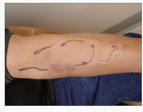
The main principle of access planning is to preserve as many veins as possible for subsequent use when the access fistula or device stops working, which inevitably will happen if the patient lives long enough. Thus, the surgeon works from the most distal location possible to more central locations, starting with the nondominant upper extremity, then the dominant upper extremity, then more unusual locations such as the femoral or axillary arteries. Native arteriovenous fistulas are used first whenever possible, then prosthetic material at each level. The order of preferred access procedures is typically a wrist or snuffbox fistula between the radial artery and the cephalic vein (the snuffbox is the space between the extensor hallucis longus and the extensor pollicis brevis at the base of the thumb, where a distal branch of the radial artery and the cephalic vein lie in close proximity), a brachial artery-to-cephalic vein fistula at the elbow, and finally a transposition of the basilic vein in the arm to the brachial artery (note that the “arm” is the proper anatomic term for what is frequently referred to as the “upper arm”). There are many variations on this theme that can be used by the creative surgeon. For example, the basilic vein in the forearm, which runs too dorsally for use in dialysis, may be transposed and connected to the radial artery, or the median antebrachial vein may be connected in a side-to-side fashion in the upper forearm to the radial artery with lysis of vein valves allowing reverse flow through this vein into the forearm venous system.7 These unusual procedures can produce complicated anatomy, which should be diagramed by the surgeon to allow the dialysis technicians and sonographers to understand the anatomy when placing the patient on dialysis or studying the fistula for possible abnormalities (Fig. 33.2).
TABLE 33.2 ADVANTAGES OF PROSTHETIC GRAFTS FOR DIALYSIS ACCESS
Large surface area/length available for cannulation
Technically easy to cannulate
Short time from insertion to maturation
Multiple insertion sites available
Variety of shapes and configurations available to facilitate placement
Easy to handle and implant
Reliably high flow
Comparatively easy to repair surgically
The main purpose of preoperative duplex scanning is to confirm the patency and determine the size of the cephalic and basilic veins in the upper extremity. Many studies have shown that the main predictor of success for a new access is the size of the outflow vein. Most authors state that a minimum diameter of 2.5 mm is required for a reasonable chance of successful maturation for a native arteriovenous fistula, although some now suggest that the failure rate is unacceptable for veins below 3 mm. A vein diameter of at least 3 mm, and preferably 4 mm or greater, is required for a prosthetic graft. In addition to having an adequate caliber, veins must be superficial enough for ease of puncture. Veins deeper than 6 mm from the skin surface are difficult to access unless they are exceptionally large. Thus, depth is an important consideration. Excessive depth does not preclude use of the vein but often requires the surgeon to “superficialize” it—surgically transposing it to a more superficial location. Patency of the central veins should also be assessed, especially in patients who have had prior access through central venous catheters.
FIGURE 33.2. A proximal radial artery arteriovenous fistula with skin markings to indicate the path of flow, which is bidirectional in this case. Such diagrams are useful for both the dialysis technicians who access the fistula and the sonographers who scan them. (Courtesy of William C. Jennings, M.D.)
A complete evaluation must include the arterial supply to the extremity. Renal failure patients are particularly prone to develop calcification of their arteries due to secondary hyperparathyroidism and an imbalance of calcium and phosphate. Heavily calcified arteries may not be suitable for access use owing to the difficulty in suturing these vessels. These patients develop occlusive disease in arteries from the aortic branches all the way to the digital vessels, which can result in inadequate flow for successful dialysis. Arterial occlusive disease also places patients at increased risk for clinically significant steal that can cause irreversible pain, loss of sensation, and weakness in the hand, as discussed later in this chapter. A preoperative digit-brachial pressure index of less than 0.6 should raise concern for the risk of steal. Some examiners check the response to hyperemia created by simple fist clenching. There should be an increase in diastolic flow with release of the clenched fist. Absence of this response suggests lack of reserve and increased risk for steal.
As described in Table 33.3, a predialysis access duplex mapping examination consists of three main components: (1) central venous mapping, (2) superficial venous mapping, and (3) arterial mapping. Specific mapping protocols should be developed at each facility by the local vascular technologists and access surgeons. Some consider an examination complete if the duplex of the nondominant arm is “normal.” Most often, the nondominant arm is used first; however, the referring physician or surgeon may want to use the patient’s dominant arm in unusual circumstances such as stroke affecting the dominant side. In general, if “right only” or “left only” is not specified, the vessels are evaluated bilaterally (this applies to the upper or lower extremities) to ensure that all possible critical information is obtained. Adequate communication with the referring physician is particularly important prior to revision of an existing dialysis access fistula or graft to make sure that questions about anatomy, outflow veins, potential substitute veins, or arterial inflow are answered.
A standard duplex ultrasound system is required for this examination with high-resolution B-mode imaging, color Doppler, and spectral waveform analysis. A selection of transducers with a choice of operating frequencies and “footprints” is necessary for scanning of the central and peripheral extremity vessels. These include a midrange-frequency linear array transducer (e.g., L7-4) for general applications and a high-frequency transducer (e.g., L12-5, L10-5 intraoperative, L15-7 intraoperative) for assessing the superficial veins. A small curved array transducer (e.g., C8-5), such as one designed for scanning neonatal heads, is excellent when scanning around the clavicle.
The examination begins with the patient in the supine position for bilateral brachial artery pressures and with the head slightly turned away from the limb being examined for evaluation of the subclavian and axillary veins. For evaluation of the arm veins, the head of the bed may be elevated 45 degrees with the arm dependent. Warming and a tourniquet, as described later in this chapter, may promote distention of the superficial veins. Several examiner positions can be utilized to scan the arms. Whenever possible, the examiner is positioned closest to the limb being evaluated (e.g., scan the right arm from the patient’s right side). Reaching across the patient’s body with one arm and operating the controls on the ultrasound system with the other arm causes muscle strain for the sonographer and can lead to musculoskeletal injuries. Thus, for a bilateral examination, after scanning the first arm, the machine may be moved to the contralateral side. Alternatively, if the examiner prefers to scan with one hand but still avoid reaching across to the contralateral side, the patient may be turned 180 degrees.
TABLE 33.3 PREDIALYSIS ACCESS MAPPING
1. Central venous mapping
Scan the innominate, subclavian, and axillary veins to evaluate for patency and flow pattern.
2. Superficial venous mapping
Scan the cephalic vein from wrist to shoulder, noting any anatomic anomalies and evidence of phlebosclerosis. Diameter should be carefully documented throughout the entire course of the vein. Map and mark the vein.
Scan the basilic vein from its origin to its confluence with the brachial vein near the axilla, noting any anatomic anomalies and evidence of phlebosclerosis. Diameter should be carefully documented throughout the entire course of the vein. Map and mark the vein.
Document patency and size of the median cubital vein. Map and mark the vein.
3. Arterial mapping
Obtain bilateral brachial systolic blood pressures and Doppler waveforms.
Scan the brachial, radial, and ulnar arteries, documenting any evidence of atherosclerosis, abnormalities, or anomalies (e.g., high bifurcation of the brachial), and any stenosis, which must be confirmed with spectral waveform analysis.
Allen’s test may be performed if the veins are acceptable.
If abnormal (digit pressure drops to <80 mm Hg or a >30% drop with compression of the radial artery), proceed to the next limb area.
If normal, measure ipsilateral first and third finger pressures (if not already done).
If no acceptable vein is found, proceed to contralateral arm.
If neither upper extremity is acceptable, proceed to lower extremity.
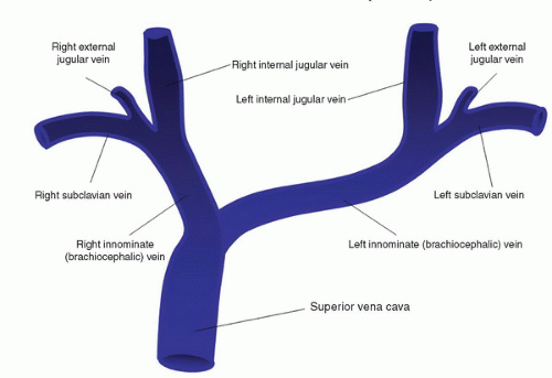
Central Venous Mapping
The normal anatomy of the upper extremity central veins is illustrated in Figure 33.3. Patency of the central veins should be ensured as far as possible, particularly in patients who have had prior venous access through puncture of these veins. Patency is compromised in about 20% of patients who have had indwelling central catheters for dialysis or other purposes. For these reasons, the central veins should be scanned first when performing a predialysis access mapping examination. The two main forms of central venous obstruction are stenosis and thrombosis. Stenosis in the innominate or subclavian vein is particularly common in patients with prior central venous catheters for short-term dialysis. Doppler findings with subclavian vein stenosis include a focal increase in Doppler flow velocity with poststenotic turbulence centrally and a continuous flow pattern peripherally. The duplex evaluation for upper extremity deep vein thrombosis is discussed in Chapter 20. Doppler waveforms from the innominate (brachiocephalic), subclavian, and axillary veins are all assessed for patency, normal flow pattern, and symmetry with the contralateral side.
The supraclavicular subclavian vein is a common location for venous stenosis where the subclavian vein curves to join the internal jugular vein. This segment often contains a valve that can become stenotic due to fibrosis of the leaflets. The infraclavicular subclavian vein is more easily imaged but is less likely to be stenotic. Doppler waveform analysis of flow in the infraclavicular subclavian vein can provide clues to the presence of a more proximal obstruction.
The axillary vein is a less common location for obstruction but may be a site of numerous collateral flow channels when a more central stenosis or occlusion exists. Subclavian vein obstruction may cause reversed flow in the axillary vein, which drains into chest wall collaterals. These prominent subcutaneous chest wall veins may be visible.
FIGURE 33.3. Anatomy of the upper extremity central veins.
Superficial Venous Mapping
The entire cephalic vein (from the wrist to its confluence with the subclavian vein) should be scanned noting any branching, wall thickening, or thrombosis (Fig. 33.4). The diameter of the cephalic vein is measured along its entire course. A tourniquet can also be useful. Some examiners advocate use of two tourniquets, one above the elbow to occlude the deep veins and one below to occlude more superficial veins. The examination may begin without the use of a tourniquet. A diameter of 3 mm or greater is generally adequate. A tourniquet should be used if vein diameter is less than 3 mm. Diameter should be recorded with a note regarding use or nonuse of a tourniquet. Vein measurements can be obtained using either a long-axis or a transverse image, with minimal pressure on the skin to avoid compressing the vein. The long-axis view has been recommended to avoid inadvertent use of an oblique transverse image, which can cause overestimation of the diameter. If the cephalic vein is inadequate, a survey should be done for other veins in the distal half of the forearm. If other forearm veins are 3 mm or larger, they should be mapped and marked from the wrist to the antecubital fossa.
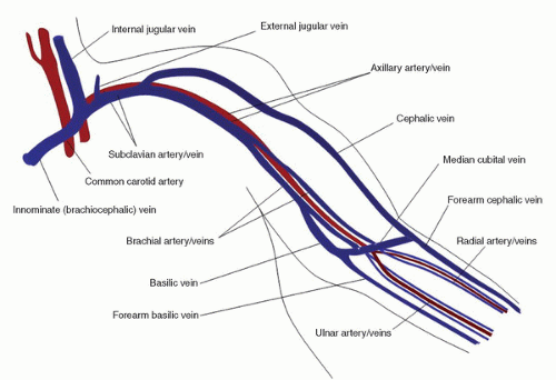
FIGURE 33.4. Anatomy of the upper extremity arteries and veins.
The basilic vein is examined from the wrist to the confluence with the brachial vein, noting the confluence location, branching, wall thickening or thrombosis, and vein diameter. Because the brachial vein is sometimes used for a transposition fistula, the diameter of this vessel should also be obtained. The anatomy of the basilic vein is quite variable; it may confluence with the brachial vein at the proximal or mid upper arm or with the axillary vein more centrally.
The median cubital is a short superficial vein that passes across the anterior aspect of the elbow between the cephalic and the basilic veins. The anatomy of the median cubital vein is extremely variable. Whenever it is associated with a patent basilic or cephalic vein, the diameter and patency of the median cubital vein should be documented.
Arterial Mapping
Bilateral brachial systolic blood pressures are obtained with the patient supine to assess for occlusive disease in the innominate, subclavian, and axillary arteries. Doppler velocity waveforms are also obtained from the brachial arteries in both arms. However, blood pressures generally should not be taken in arms with a patent fistula or graft, to avoid inadvertent thrombosis of the access. The subclavian and axillary arteries are scanned, with particular attention to these segments if the brachial systolic blood pressures are unequal. Normal velocity waveforms in the upper extremity arteries are triphasic with no significant focal velocity increases. Upper extremity arterial duplex scanning is discussed in Chapter 14. The brachial, radial, and ulnar arteries are scanned throughout their entire course, noting any atherosclerosis, tortuosity, medial calcification, or anatomic variants such as a high bifurcation of the brachial artery into the radial and ulnar branches in the proximal arm.
Palmar Arch (Allen’s Test)
If the forearm arteries and veins appear adequate for creation of a forearm fistula or graft, the palmar arch is evaluated to determine whether the blood supply to the hand is dependent on either the radial or the ulnar artery alone. The ulnar artery is often the dominant of the two forearm arteries and, therefore, is usually preserved for arterial supply to the hand. The radial artery is often nondominant and can be utilized for arterial inflow to the access site with little risk of hand ischemia. Patency of the palmar arch, which connects the radial and ulnar artery circulations, is evaluated with Allen’s test. In the classic, less reliable form, this test uses simple observation of the return of pink color to the palm after clenching the fist with either the radial or the ulnar artery manually compressed. Slow return of color with one or the other artery manually occluded suggests that the hand is reliant on the occluded artery for its primary blood supply. A more quantitative version of Allen’s test can be performed using photoplethysmography (PPG), as outlined in Table 33.4. Duplex scanning of the palmar arch can also be used for a direct evaluation of flow from the radial and ulnar arteries into the palm, using manual occlusion of these arteries at the wrist. These variations of Allen’s test are described in more detail in Chapter 34.
Only gold members can continue reading. Log In or Register to continue