Diagnostic Catheter-Based Vascular Angiography


Diagnostic Catheter-Based Vascular Angiography


Debabrata Mukherjee


Angiography allows direct visualization of blood vessels in the body by the injection of iodinated contrast material via a catheter placed directly into the artery or vein. It remains the gold standard to determine the severity and extent of peripheral arterial disease. Digital subtraction angiographic technology allows high-quality images using small amounts of contrast material. The field of angiography has grown tremendously in recent years, and today we have percutaneous methods for accessing virtually every artery in the human body. A number of unique catheter shapes have been developed to facilitate intubation/cannulation of the ostium of specific arteries. However, angiography is invasive and is typically indicated only in patients in whom revascularization is being considered.


ANATOMIC CONSIDERATIONS


Access for Vascular Angiography

The choice of the appropriate arterial access site is crucial. Typically for the purposes of endovascular intervention, the access site should be as close to the intended lesion as possible. The location of the lesion determines the access site in most cases, and approach to iliac occlusive disease is most commonly made from the ipsilateral femoral artery via retrograde common femoral artery (CFA) access. Lesions of the CFA, however, must be approached from a contralateral-puncture, axillary, or popliteal approach. Lesions distal to the CFA are best approached and treated with an antegrade CFA approach. For renal, mesenteric, and cerebral angiography, the typical access site is retrograde CFA.


Retrograde Common Femoral Artery Access

This is the commonest access site used for diagnostic angiography. The CFA is palpated below the inguinal ligament, which courses between the pubis and the anterior superior iliac spine. In patients with peripheral arterial disease, one should always check the site/level under fluoroscopy prior to arterial puncture. In individuals with severe peripheral arterial disease, the femoral pulse is often not palpable, and in those instances, ultrasoundguided approach should be used or an alternative access site should be used. The SmartNeedle (Escalon Vascular Access Inc., New Berlin, WI) is a percutaneous Doppler-guided vascular access device, which uses a handheld monitor and a Doppler transducer located at the tip of an access needle to provide continuous auditory feedback and is often helpful in patients with absent or weak pulses.


Brachial/Radial Artery Access

Brachial artery access is considered in individuals with bilateral iliac artery or distal aortic occlusions. In some individuals with severely angulated origin of the visceral vessels, that is, celiac, superior mesenteric, or renal arteries, brachial approach may make selective cannulation easier. Left brachial approach is preferable to minimize the potential risk of stroke, as entry from the right exposes both the carotid and vertebral arteries to risk of embolization. The risk of injury and thrombosis is significantly higher with brachial compared to femoral access. It is recommended that the operator injects 3,000 to 5,000 units of unfractionated heparin in the sheath after brachial/radial access to minimize risk of thrombosis. If the radial artery is used for access, 50 to 100 µg of intra-arterial nitroglycerin or verapamil is administered to minimize spasm in addition to heparin.


Popliteal Artery Access

In individuals with occluded superficial femoral arteries, the popliteal artery may be cannulated for diagnostic angiography. Limitations to this access site include the requirement that the patient lies prone during the procedure and the difficulty in localizing the artery. Doppler-guided needle approach should ideally be used to access the popliteal artery. This access site is rarely used for diagnostic angiography and more commonly used for interventions.


FUNDAMENTALS

Angiography involves direct injection of iodinated contrast material into the blood vessels for their visualization. Many different contrast agents are available today for angiography. The two clinically important attributes of a contrast agent are its iodine dose and osmolality. To maintain good radiographic efficacy and safety, contrast agents must balance the somewhat paradoxical relationship between these two properties. Iodine dose refers to the amount of iodine delivered in an injected dose of contrast material. The iodine, delivered by iodinated benzene ring compounds, produces radiographic “contrast” by blocking x-rays. Visualization is typically improved by increasing the iodine load, a function of the percentage of iodine and the concentration of the compound present upon injection. Increasing the iodine load, however, results in increased osmolality. Osmolality refers to the number of dissolved particles in a solution or the concentration. Ideally, contrast agents injected into the vasculature should have an osmolality as close to that of body fluids as possible. Solutions with osmolality greater (hypertonic) or less (hypotonic) than that of body fluids can cause cells to shrink or swell, respectively, contributing to numerous hemodynamic, physiologic, and biologic adverse effects. The body also attempts to quickly dilute and excrete hypertonic solutions to maintain osmotic equilibrium. Therefore, the benefits gained from increasing the iodine load in contrast agents to improve radiographic efficacy may be offset by the adverse effects associated with higher osmolality solutions.
The goal should be to use the lowest dose and volume of contrast necessary for adequate clinical angiography. Broadly, there are two types of agents, high-osmolality and low-osmolality agents. The major resistance to the use of low-osmolality agents used to be expense, but recent price reductions have made this a relative nonissue. High-osmolality agents are rarely used in practice now. Third-generation nonionic contrast agents reduce osmolality even further by creating a dimer. Iodixanol is a dimeric contrast agent in this class and is iso-osmolal with plasma. Nonionic, low osmolal contrast agents are now routinely used for angiography. Individuals with prior significant contrast reactions, active asthma, severe congestive heart failure, active significant arrhythmia, aortic stenosis, pulmonary hypertension, or significant renal dysfunction (serum creatinine greater than 2 mg/dL) should receive a low-osmolal or preferably an iso-osmolal agent. For diagnostic vascular angiography, we use an iso-osmolal agent iodixanol (Visipaque, GE Healthcare, Little Chalfont, Buckinghamshire) that has been shown to cause significantly less discomfort for patients and also significantly reduces the relative risk of developing contrast media-induced renal failure. In individuals with prior severe allergic reaction to contrast agents, gadolinium can be used for angiography using digital subtraction angiography (DSA) provided GFR is greater than 30 mL/min. Carbon dioxide has a limited role as a vascular contrast agent because of poor opacification of the vessels despite use of DSA.


CLINICAL ASPECTS OF ANGIOGRAPHY

Knowledge of appropriate catheters, angulations, and injection rates is imperative in performing safe and optimal vascular angiography. Table 3.1 makes general recommendation for catheter selection and injection rates in different vascular territories.


Lower Extremity Angiography

Despite recent advances in the noninvasive evaluation of lower extremity vascular disease, contrast angiography remains the gold standard. A pelvic/abdominal aortogram (Fig. 3.1) is initially performed with a straight pigtail catheter (Fig. 3.2) placed at the level of L1-L2 slightly above the level of the aortoiliac bifurcation (typically L3). This allows excellent visualization of the distal aorta and the origin of the common iliac arteries and the external iliac and the common femoral vessels. Angulated views are indicated to visualize the iliac and the femoral bifurcations without overlap. A left anterior oblique (LAO 30 degrees) view allows visualization of the left common iliac and right common femoral bifurcations without overlap. Digital subtraction pelvic aortograms are performed with 10 to 15 mL of iso-osmolal contrast at a rate of injection of 15 mL/s. Following the pelvic aortogram, the pigtail catheter is withdrawn to the aortic bifurcation so that the injected contrast fills the runoff vessels bilaterally with minimal contrast diverted to the viscera. DSA with moving table and bolus chase technology to visualize the outflow (Fig. 3.3A) and the runoff vessels


is recommended (Fig. 3.3B). The bolus chase technology combines the advantages of digital subtraction and step table technology. Mask images are obtained prior to acquisition of contrast images to further accentuate the arterial tree. Figure 3.4 is a schematic of the arterial supply to the lower extremities.


TABLE 3.1 RECOMMENDATIONS FOR DIAGNOSTIC VASCULAR ANGIOGRAPHY


Artery


Catheter


Angulation


Injection


Arch aortogram


5 FR angulated pigtail at aortic root


30° LAO


10 mL/s for 3 s


Abdominal aortogram for suspected mesenteric ischemia


5 FR straight pigtail between T12 and L1


Biplane or lateral


20 mL/s for 2 s


Abdominal aortogram for suspected renal artery stenosis


5 FR straight pigtail between T12 and L1


AP


20 mL/s for 0.5 s using DSA


Pelvic/abdominal aortogram


5 FR straight pigtail between L1 and L2


AP


15 mL/s for 1 s using DSA


Distal aorta for bolus chase and runoffa


5 FR straight pigtail between L2 and L3


AP


8 mL/s for 10 s using DSA


Carotids and great vessels


JR4 catheter for level 1 arch


Ipsilateral oblique 30° and lateral


Hand injection with DSA


Vitek or Headhunter catheter for level 2 arch


Ipsilateral oblique 30° and lateral


Hand injection with DSA


Simmons 1 or 2 for level 3 arch


Ipsilateral oblique 30° and lateral


Hand injection with DSA


Renal arteries


JR4 catheter, SOS catheter


AP and ipsilateral oblique at 20°-30°


Hand injection with DSA


Celiac trunk


JR4, SOS, Cobra catheters


AP


Injection at 10 mL/s


Superior mesenteric artery


JR4, SOS, Cobra catheters


AP


Injection at 8 mL/s


Inferior mesenteric artery


JR4, SOS, Cobra catheters


AP


Injection at 3 mL/s


Pulmonary artery


Grollman catheter


LAO/RAO 30°


20 mL/s for 2 s


a The volume should be reduced by 50% if each leg is injected separately.


LAO, left anterior oblique; RAO, right anterior oblique; AP, anteroposterior; DSA, digital subtraction angiography.


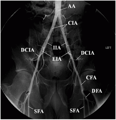
FIGURE 3-1. Pelvic aortogram showing common iliac arteries and their bifurcation. Common femoral artery and its bifurcation are also displayed. (AA, abdominal aorta; CIA, common iliac artery; EIA, external iliac artery; IIA, internal iliac artery; DCIA, deep circumflex iliac artery; CFA, common femoral artery; SFA, superficial femoral artery; DFA, deep femoral artery)


FIGURE 3-2. A straight pigtail catheter (Omniflush) is used to perform abdominal and pelvic aortograms.


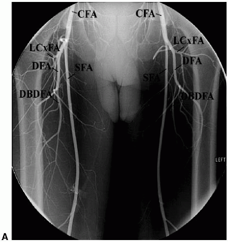
FIGURE 3-3. A: Bilateral lower extremity outflow angiography; anteroposterior view with digital subtraction technique. (CFA, common femoral artery; DFA, deep femoral artery; SFA, superficial femoral artery; LCxFA, lateral circumflex femoral artery; DBDFA, descending branch of deep femoral artery) (Continued)

Jun 12, 2016 | Posted by in CARDIOLOGY | Comments Off on Diagnostic Catheter-Based Vascular Angiography

Full access? Get Clinical Tree

Get Clinical Tree app for offline access