Chapter 39 Diagnosis and Management of Asthma in Adults
Asthma is a disorder of the airways that is characterized by typical symptoms arising from a complex interplay between chronic inflammation and disordered airway function. Worldwide disease prevalence has, until recently, risen steadily, and the condition contributes to a significant amount of morbidity and is responsible for many preventable deaths, particularly in developed countries, where 1 in 10 children and 1 in 20 adults have a diagnosis of asthma. The goals of management of asthma are to make an accurate diagnosis; to quantify current morbidity and assess risk of future morbidity; and to use pharmaceutical and nonpharmaceutical interventions to eliminate or minimize current symptoms and future risk of asthma attacks and accelerated decline in lung function. Because asthma usually is a lifelong disease, good patient education and a collaborative approach to management can be expected to increase the chances of success. Pharmacologic management involves the stepwise use of β-agonist bronchodilators, inhaled corticosteroids, and other agents in dosages usually titrated according to symptom management. Important nonpharmacologic measures include patient education, avoidance of triggers, and smoking cessation. Satisfactory control of asthma is achieved in a majority of patients. However, between 5% and 10% of cases of so-called refractory asthma remain poorly controlled and contribute disproportionately to asthma-related morbidity, health care costs, and mortality. The reasons for this are complex and multifactorial, and many patients with refractory asthma require referral to specialist centers.
Definition and Key Features of Asthma
Asthma is derived from the Greek word aazein, meaning “to labor in breathing” and was first used by Hippocrates, in 450 BCE, to describe a condition characterized by spasms of breathlessness. The present Global Initiative for Asthma (GINA) definition of the disease (Box 39-1) is a lengthy description of histopathologic, pathophysiologic, and clinical features that encompass the major disease characteristics. Fundamental features are airway hyperresponsiveness, chronic airway inflammation, disordered airway mucosal immunity, and structural changes to the airways (airway remodeling).
Box 39-1
Global Initiative for Asthma (GINA) Description of Asthma
Asthma is a chronic inflammatory disorder of the airways in which many cells and cellular elements play a role, in particular, mast cells, eosinophils, T lymphocytes, neutrophils, and epithelial cells. In susceptible individuals, this inflammation causes recurrent episodes of wheezing, breathlessness, chest tightness, and cough, particularly at night and in the early morning. These episodes are usually associated with widespread airflow obstruction that is typically reversible either spontaneously or with treatment.
From Global Strategy for Asthma Management and Prevention (Update 2009). Available at www.ginaaasthma.org.
Airway Hyperresponsiveness
Airway hyperresponsiveness is considered to be the cardinal pathophysiologic abnormality in asthma. It represents an exaggerated bronchoconstrictor response to a variety of largely exogenous inhaled stimuli causing bronchoconstriction, either by a direct effect on airway smooth muscle or indirectly by interacting with neural pathways or mast cells. Airway hyperresponsiveness is likely to be the basis for the variable airflow obstruction that is responsible for many of the day-to-day symptoms of asthma, including those associated with exercise-induced asthma, nocturnal asthma, and asthma induced by fumes or cold air.
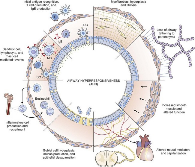
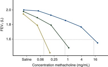
Airway hyperresponsiveness can be objectively demonstrated as a 20% fall in the forced expiratory volume in 1 second (FEV1) after inhalation of histamine or methacholine) at a concentration below 8 mg/mL. This represents an abnormal effector response of airway smooth muscle, characterized by heightened pharmacologic sensitivity and reactivity to the bronchoconstrictor stimulus (Figure 39-1). Naturally occurring airway hyperresponsiveness reflects an abnormally amplified response of airway nerves and mast cells to exogenous stimuli, as well as an intrinsic abnormality of the airway smooth muscle response. The basis for this generalized hyperresponsiveness is not entirely clear, but sensitization of airway nerves, mast cells, and smooth muscle by inflammatory mediators, along with loss of epithelial barrier function, reduced production of bronchoprotective factors, an intrinsic abnormality of airway smooth muscle, and structural changes to the airway, all are likely to play a part (Figure 39-2). One key pathologic feature associated with airway hyperresponsiveness in asthma not seen in nonasthmatic eosinophilic bronchitis is infiltration of the bronchial smooth muscle layer by mast cells, implying that the interaction between these cells is fundamentally important in the pathogenesis of airway hyperresponsiveness.

Figure 39-1 Airway responsiveness measured as the fall in forced expiratory volume in 1 second (FEV1) after increasing inhaled concentrations of methacholine. Severe (yellow), moderate (green), and mild (blue) airway hyperresponsiveness are shown. Note increased airway responsiveness is associated with a lower provocative concentration required to cause a 20% fall in FEV1 (PC20) (see green line), a steeper gradient, and a higher maximum % fall in FEV1.
Chronic Airway Inflammation
Histopathologic examination of postmortem specimens from patients with fatal asthma show an inflammatory response characterized in many cases by the presence of airway eosinophilia. Typically, eosinophilic infiltration can be found throughout the airway wall, within thick viscid plugs that occlude the airway lumen and often extend into the lung parenchyma and alveolar spaces and even into adjacent blood vessels (see Figure 39-2). In addition, extensive eosinophilic degranulation with deposition of major basic proteins occurs. Associated findings include widespread shedding of the airway surface epithelium, thickening of the reticular basement membrane, and enlargement of airway smooth muscle and submucosal glands. A minority of patients dying from asthma, particularly those with sudden-onset fatal asthma, exhibit evidence of eosinophilic inflammation. In these cases it is believed that widespread mast cell degranulation is the primary event with evidence for a relative excess of neutrophils in the distal airways and parenchyma.
The limited number of patients studied by bronchoscopy makes it difficult to investigate heterogeneity of the lower airway inflammatory response, but less invasive assessment of airway inflammation using induced sputum analysis has shown that a significant proportion of patients with asthma studied when stable and during an attack have normal eosinophil numbers in induced sputum samples. This sputum cell profile has been reported in patients with severe asthma and in patients who are not treated with inhaled corticosteroids, and the absence of eosinophilic airway inflammation has been confirmed by bronchoscopy studies. Thus, the presence of a distinct noneosinophilic corticosteroid-resistant asthma phenotype across the range of asthma severity seems secure. Noneosinophilic asthma is discussed in more detail later in the chapter.
Disordered Airway Mucosal Immunity
The airway mucosa in persons with asthma mounts an abnormally amplified immunologic response to a variety of exogenous stimuli, including inhaled allergens, cigarette smoke, infectious agents, and air pollutants. Some or all of these abnormal responses may be the consequence of failure of maturation of the immune response in early life as a result of reduced exposure to pathogens at a critical point in development.
Particular interest has focused in the response to allergen, because atopy is twice as common in patients with asthma as in control subjects without asthma, and inhalation of allergen in a patient with atopic asthma who is sensitized to the allergen results in a marked eosinophilic airway inflammatory response, developing 4 to 6 hours after inhalation associated with airflow obstruction (the late response) and increased airway responsiveness lasting for days and weeks. Common aeroallergens in many Westernized countries include house dust mite, cat fur, grass pollen, ragweed, and Aspergillus fumigatus spores. Most patients with childhood-onset asthma are sensitized to one of more of these allergens, and many have extrapulmonary manifestations of allergy, including eczema and allergic rhinitis. Opinions regarding the significance of the response to allergen vary, ranging from a fundamentally important role in the pathogenesis of airway inflammation and dysfunction in asthma to a position in which it is seen as a more peripheral mechanism. The occurrence of histopathologically similar forms of asthma in nonatopic patients and the disappointing therapeutic effect of allergen avoidance and anti-IgE treatment suggest that the latter position is correct, although uncertainty remains.
Growing evidence points to an abnormal airway response to infecting respiratory viruses, resulting in an amplified airway inflammatory response and more pronounced clinical consequences. The molecular mechanisms of this remain uncertain but constitute an active area of current study. There is little doubt that viral infection is an important trigger for acute severe asthma. Smoking and perhaps exposure to other environmental pollutants have been linked to worsening of symptoms, an increased risk of asthma attacks, and the development of fixed airflow obstruction. This association may reflect an abnormality of the innate immune response; patients with asthma who smoke or are exposed to environmental pollutants tend to have noneosinophilic, neutrophilic airway inflammation.
Structural Changes to the Airway
Structural changes in airway morphology (airway remodeling) probably occur as a result of chronic airway inflammation and dysfunction. These changes are likely to be the basis for the accelerated lung function decline and fixed airflow obstruction seen in some patients with asthma. Key features of airway remodeling include thickening of the subepithelial basement membrane caused by abnormal deposition of collagen, increased airway smooth muscle bulk, increased mucus-secreting cells, and increased airway vascularity (see Figure 39-2). In severe asthma, bronchiectasis, small airway fibrosis, and emphysema may be features. The bronchiectasis is associated with sensitization to Aspergillus.
Pathophysiologic Basis of Asthma
Asthma is a complex condition in which it is not always obvious which of the various pathophysiologic abnormalities is responsible for morbidity at any one time. In order to make some sense of this complexity, it can be helpful to consider five potentially important pathophysiologic factors, presented in alphabetical array for convenience:
These abnormalities are likely to be linked, but the mechanism is complex, and cross-sectional and longitudinal correspondence among them is not close. Accordingly, each is best considered as a relatively independent factor.
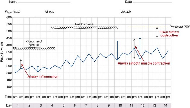
It is likely that these factors contribute to airflow obstruction and morbidity in different ways (Figure 39-3). Airway hyperresponsiveness is responsible for short-term, bronchodilator-responsive variable airflow obstruction which is the basis for many of the day-to-day symptoms experienced by patients and is suppressed by bronchodilator therapy, and to a lesser extent corticosteroids. Bronchitis, which may be eosinophilic and corticosteroid-responsive or neutrophilic and corticosteroid-unresponsive, manifests with cough and sputum and a relatively bronchodilator-resistant but potentially corticosteroid-responsive airflow obstruction of more gradual onset. Inflammation-mediated airflow obstruction is particularly important in the pathogenesis of asthma attacks. Cough and a small amount of sputum production is common in asthma. It probably reflects airway inflammation but also may be due to cough reflex hypersensitivity, a common but poorly understood and difficult-to-treat aspect of asthma and other airway diseases. Airway damage may manifest with disability caused by bronchodilator and corticosteroid-resistant airflow obstruction or mucus retention and infection as a result of airway and lung parenchymal damage. Extrapulmonary conditions linked to the inflammatory airway disease or independent of it also may contribute to symptoms (Box 39-2).

Figure 39-3 Peak expiratory flow (PEF) chart illustrating airflow obstruction due to at least three discrete mechanisms: airway smooth muscle contraction (note good acute response to inhaled salbutamol); airway inflammation (note loss of acute bronchodilator response but gradual increase in PEF with oral prednisolone); and airway damage (bronchodilator and steroid-unresponsive airflow obstruction).
Asthma Heterogeneity and Clinical Subgroups of Patients with Asthma
Asthma has long been recognized as a heterogeneous disease. As early as 1918, Francis Rackemann identified clear clinical subgroups on the basis of history, skin tests, and response to “a clinical experiment such as a change in residence, a restriction in diet or an elimination of some supposedly offending substance.” He made a distinction between “extrinsic asthma” due to “hypersensitivity to some foreign substance outside of the body and “intrinsic asthma . . . implying the essential cause of the trouble is inside of the body.” The subdivision of extrinsic (atopic) and intrinsic (nonatopic) asthma remains popular. The most obvious difference is that the peak age at onset of atopic asthma is in childhood, whereas nonatopic asthma often manifests first in adults. It has not been possible to identify consistent histopathologic differences. In general, patients with atopic asthma exhibit more airway hyperresponsiveness and better responses to inhaled corticosteroids than those with nonatopic asthma. Nonatopic asthma is associated with greater heterogeneity of the lower airway inflammatory response, because a majority of patients with noneosinophilic asthma are nonatopic.
Asthma also has been classified according to the dominant clinical characteristic. Exercise-induced asthma, premenstrual asthma, and seasonal asthma are examples. These subdivisions help to remind the clinician of the dominant trigger but do not identify patients with a distinct pathologic process or treatment response. Important exceptions are aspirin-induced asthma, asthma in endurance athletes, and occupational asthma. These conditions are considered later.
Current interest is in categorization of asthma by the pattern of lower airway inflammation. The development of induced sputum as a means to non-invasively assess airway inflammation has made it possible to do this. Cross-sectional studies show that 20% to 40% of patients with symptomatic asthma do not have sputum evidence of eosinophilic airway inflammation. Many have a sputum neutrophilia and evidence of airway release of cytokines linked to the innate immune response. This sputum profile is evident in corticosteroid-naive as well as corticosteroid-treated persons and is consistently seen in patients with asthma, suggesting it is not always an artifact related to infection or treatment. Noneosinophilic asthma is clinically important, because response to corticosteroids is less pronounced in patients with this inflammatory profile than in persons with more typical sputum features. Noneosinophilic asthma has been associated with smoking, obesity, high-level endurance training in athletes, menopause, occupational endotoxin exposure, and recurrent bacterial bronchitis. Treatment and management approaches have not been investigated extensively, but aggressive corticosteroid therapy is unlikely to be helpful. Preliminary evidence indicates that long-term macrolide antibiotics may be of benefit.
Cross-sectional sputum studies also have shown that some patients with cough have eosinophilic airway inflammation but normal airway function (eosinophilic bronchitis). Eosinophilic bronchitis is closely related to “atopic cough,” a condition described in Japan and characterized by cough, atopy, and evidence of large airway eosinophilic inflammation. Cough variant asthma initially was described as asthma manifesting with a bronchodilator-responsive chronic cough. More recently, this entity has been extended to encompass patients presenting with cough and objective evidence of asthma (i.e., variable airflow obstruction and/or airway hyperresponsiveness). All of these conditions are characterized clinically by a corticosteroid-responsive chronic cough.
Genetics of Asthma
Asthma has long been known to run in families. Studies of monozygotic and dizygotic twins, which control for environmental exposure, have estimated heritability of 30% to 70%. Considerable effort and resources have been put into identifying the genetic basis of asthma. This work is inevitably compromised by the rather general and imprecise definition of the disease, and results have been mixed and often are inconsistent. A better understanding of different phenotypes of asthma and a stronger focus on aspects of the disease (i.e., fixed airflow obstruction, airway hyperresponsiveness, atopy, eosinophilic airway inflammation), rather than on the syndrome, may be the way forward. Despite these limitations, some consistent genetic associations have been identified.
Functionally important polymorphisms of the prostaglandin D2 (PGD2) receptor gene have been associated with asthma in case control studies in racially diverse populations. PGD2 is an important product of mast cells, and the receptor is involved in T cell recruitment. Mice deficient in this gene do not develop airway inflammation in response to allergen. Interest in this receptor has increased recently, because preliminary evidence indicates that PGD2 receptor antagonists have useful therapeutic effects in asthma. Positional cloning studies have identified a consistent association between asthma and multiple single nucleotide polymorphisms of the ADAM33 gene, particularly when asthma is associated with airway hyperresponsiveness or fixed airflow obstruction. This gene may be involved in control of airway structure and myogenesis. The minor allele (G) of the functional variant of the promoter region of the matrix metalloproteinase 12 gene (MMP12) has been associated with better lung function in patients with asthma and in smokers, suggesting a role for MMP12 in maintaining lung function and raising the possibility of a common mechanism for the development of fixed airflow obstruction in smokers and in patients with asthma. Polymorphisms of the genes for IL-13 and FcεR1B, which modifies the activity of the high-affinity IgE receptor, may have a closer link with atopy, IgE, eosinophilic airway inflammation, and mucus production. Other genetic associations with asthma such as polymorphisms of the gene for the microbial pattern recognition receptors CD14, Toll-like receptor-1 (TLR-1), and this should be T cell immunoglobulin mucin-like domain-1 (TIM-1) may operate primarily by modifying innate immunity and the maturation of the immune system.
Genetic polymorphisms also may potentially influence the response to treatment. Variation in the gene for the β2-adrenergic receptor (ADRB2) has been the subject of much interest, because persons who are Gly16 homozygotes or heterozygotes have a diminished acute bronchodilator response compared with Arg16 homozygotes. On the other hand, Arg16 homozygotes are more susceptible to β2-receptor downregulation and may potentially be at risk for an adverse response to regular use of β2-agonists. Evidence for such an effect, however, has not been seen consistently, particularly with long-acting β2-agonists. Important genetic diversity in the response to corticosteroids has been more difficult to establish, in part because the response to this treatment is more difficult to quantify. The response to leukotriene antagonists has been associated with the C allele of the LTC4 synthase gene promoter (A-444C) polymorphism. This polymorphism is also associated with aspirin-induced asthma, an asthma variant associated with increased airway production of cysteinyl leukotrienes.
Diagnosis
One of the problems facing clinicians and epidemiologists is the absence of a “gold standard” modality for defining or diagnosing asthma. Characteristic clinical features (Box 39-3) coupled with objective demonstration of variable airflow obstruction and/or airway hyperresponsiveness usually will provide sufficient evidence to make the diagnosis.
Box 39-3
Factors Affecting Likelihood of Asthma as Cause of Respiratory Symptoms*
Factors Associated With Increased Probability of Asthma
More than one of the following symptoms: cough, breathlessness, wheeze, chest tightness
Symptoms worse at night and in the early morning
Symptoms occurring in response to exercise, allergen exposure, and cold air
Symptoms developing after taking aspirin or beta blockers
Family history of asthma and/or atopic disorder
Variable wheeze heard on auscultation of the chest
Variable PEF (see Clinical Examination Section)
Factors Associated With Lower Probability of Asthma
Prominent dizziness, light-headedness, peripheral tingling
Repeatedly normal physical examination of chest when symptomatic
Significant smoking history (i.e., more than 20 pack-years)
History
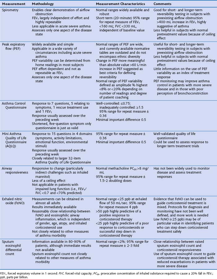
Asthma symptoms typically are variable reflecting variability in airflow obstruction and other aspects of the disease. Exaggerated diurnal variation in physiologic bronchomotor tone causes airflow obstruction and symptoms at night, maximally at 4 AM. Other symptoms reflect airway hyperresponsiveness to a multitude of possible trigger factors. Symptoms occurring after exercise, after exposure to allergens, and in response to inhaled noxious fumes such as cigarette smoke are most commonly reported. Box 39-3 lists some of the symptoms that increase and decrease the probability of asthma. An assessment of disease severity can be made from the frequency of daily symptoms, exercise limitation, nocturnal wakening, and exacerbation frequency (Table 39-1).
The pattern of daytime symptoms and seasonal variations can help identify common triggers. In patients with adult-onset symptoms, the possibility of occupational asthma or an alternative diagnosis should be carefully explored. Symptoms that worsen at work but remit outside of the workplace should alert the clinician to the possibility of occupational asthma.
Clinical Examination
Findings on the clinical examination frequently are normal in persons with well-controlled symptoms. Patients with persistent symptoms may display features of obstructive airway disease, notably a hyperinflated and hyperresonant chest and diffuse polyphonic expiratory wheeze. These signs are indistinguishable from those found in chronic obstructive pulmonary disease (COPD). Physical examination is helpful for identifying features of an alternative diagnosis.
Measuring Airflow Obstruction
The demonstration of obstructive spirometry in patients with a history suggestive of asthma strongly supports the diagnosis and is a strong basis to commence therapy. Spirometry is the preferred method for demonstrating airflow obstruction, because it is less effort-dependent than peak expiratory flow. Airflow obstruction is defined as an FEV1/FVC ratio of less than 0.7.
Measuring Variability in Airflow Obstruction
The spirogram frequently is normal in patients with asthma who are asymptomatic at the time of testing. In this clinical scenario, measurement of variability in airflow obstruction will provide useful additional information. A number of different methods exist to measure variability:
Diurnal variation in peak expiratory flow (PEF): Calculated from serial PEF measurements (see Figure 39-3); typically expressed as the difference between the highest and lowest daily readings as a percentage of the mean (see Table 39-1)
Assessment of the bronchodilator response: Measured as the change (improvement) in FEV1 or PEF from baseline with either a short-acting bronchodilator (bronchodilator responsiveness) or after a therapeutic trial of corticosteroid (steroid responsiveness)
Assessment of airway responsiveness: Measuring the fall in FEV1 in response to bronchoconstrictor stimuli, including pharmacologic agents (methacholine or histamine challenge tests), exercise, and allergen challenge (see Figure 39-1)
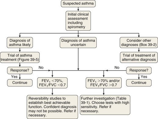
The measurement characteristics of the various tests to assess variable airflow obstruction and asthma in general are outlined in Table 39-1. An algorithm for the assessment of patients with suspected asthma is presented in Figure 39-4.
Diagnostic Pathway
The diagnostic pathway followed will depend on the pretest probability for the diagnosis, response to therapy, and level of clinical suspicion for an alternative diagnosis. The scope of the differential diagnosis is quite different in patients with and without airflow obstruction (see Box 39-2), and the main diagnostic question also is different. In the former, the clinician often is seeking support for a clinical diagnosis of asthma and the initiation of antiasthma therapy; in the latter, the evidence for an airway problem is more clear-cut, and the question is not whether inhalers should be used but how intensive the corticosteroid component of that therapy should be.
Vocal cord or glottic dysfunction is an important condition that can cause serious diagnostic difficulty in both adolescents and adults and, if not recognized, unnecessary overtreatment. Wheezing that arises from the glottis is heard throughout the lung fields but also is easy to hear without the stethoscope, with prominent noises arising from the neck. Direct visualization of the glottis by laryngoscopy may reveal the characteristic inspiratory apposition of the cords. Sometimes persons who have asthma make this noise, because they subconsciously feel the need to impress on the examiner the severity of their condition. In other patients, the noise occurs for purely psychological reasons, and no evidence of asthma is found. Patients may be of either gender but are often women in the age range of 16 to 50 years, who may have a paramedical background. Their “asthma” seems “resistant” to standard treatments, and often they have been hospitalized on many occasions and been treated unsuccessfully with large doses of corticosteroids and other treatments. Home peak flow readings and attempts at spirometry may be variable but show little correlation with attacks or treatment. Flow-volume curves may show a characteristic “fluttering” of the inspiratory curve. Measurement of total airway resistance in a body plethysmograph may be diagnostic, because the panting maneuver necessary for such measurement abolishes the vocal cord adduction, and airway resistance can be shown to be normal. Occasionally patients end up being mechanically ventilated because of “severe” asthma, but once pharmacologic paralysis is achieved, it can be seen that airway resistance is normal and that there is no necessity for high inflation pressures.
Vocal cord dysfunction probably is much more common than has been appreciated and if the condition is underdiagnosed or the relevant symptoms are mistaken for asthma, overtreatment is likely. Correct diagnosis is essential, and if vocal cord dysfunction is the sole or major part of the wheezing disorder, speech therapy can be helpful.
Diagnostic Strategy in Patients With Normal Spirometry Findings
In patients with normal spirometry findings, the primary considerations in the differential diagnosis are non–bronchodilator-, non–corticosteroid-responsive conditions. All tests of variable airflow obstruction lack sensitivity in this setting, meaning that negative findings are not particularly helpful. Positive findings are obtained more frequently with repeated testing or if care is taken to perform the assessment when the patient is symptomatic. Assessment of airway hyperresponsiveness is a more sensitive test, so the absence of airway hyperresponsiveness should prompt careful consideration of an alternative diagnosis.
Diagnostic Strategy in Patients With Obstructive Spirometry
In patients with obstructive spirometry, tests of airway responsiveness and reactivity are less specific for a diagnosis of asthma. The measurement of airway hyperresponsiveness is therefore not helpful. Bronchodilator challenge tests and therapeutic trials of corticosteroid treatment may aid diagnosis, because larger changes in FEV1 (i.e., more than 400 mL) are very suggestive of asthma. In many cases it may not be possible to assign a precise diagnostic label, although bronchodilator and steroid reversibility studies do help guide the intensity of therapy with these agents.

Full access? Get Clinical Tree