Diabetes, Hypertension, and Hypercholesterolemia
A 54-year-old man has diabetes, hypertension, and hypercholesterolemia. He complains of dyspnea on exertion and has limited his activities.
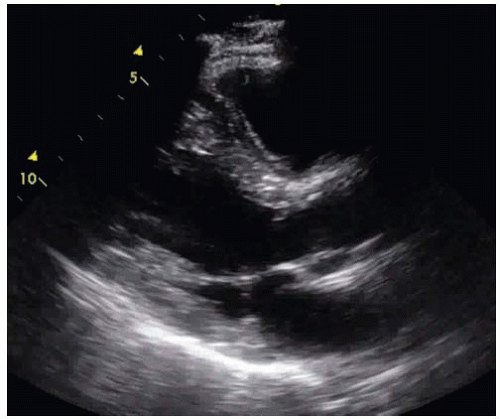
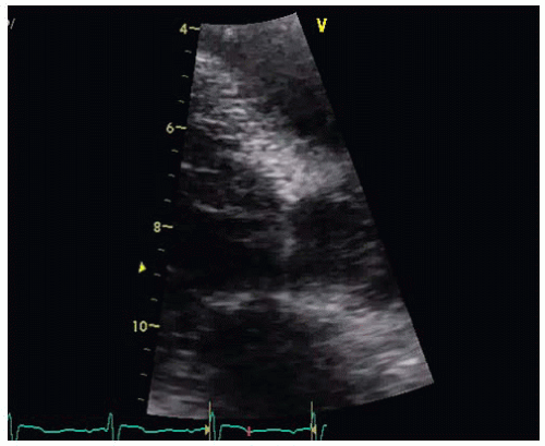
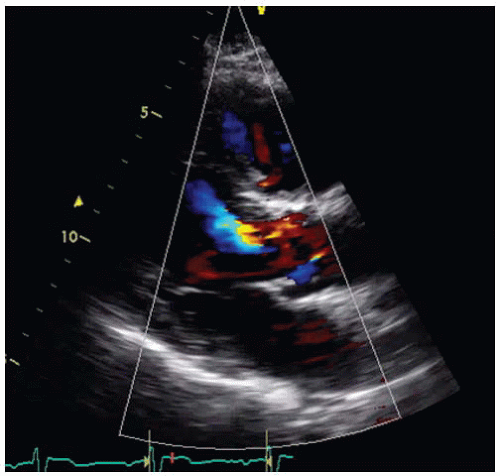
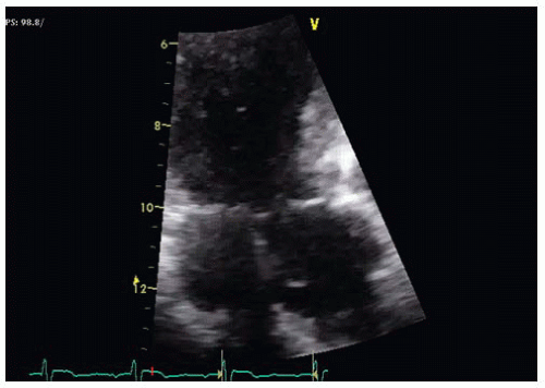
A stress test showed no evidence of inducible ischemia. He has a complete 2D, color, and Doppler transthoracic echocardiogram (Figs. 36-1, 36-2, 36-3 and 36-4 and Videos 36-1 to 36-6).
QUESTION 1. The patient’s dyspnea may be due to:
A. Severe aortic valve stenosis
B. Significant mitral regurgitation
C. Dilated cardiomyopathy
D. A subaortic membrane
E. Aortic regurgitation
View Answer


ANSWER 1: D. The patient has a subaortic membrane. The images illustrate the importance of thorough interrogation of the area of the left ventricular outflow tract (LVOT) and implementing use of color Doppler. Figure 36-1 does not visualize any aortic valve disease or a membrane. Figure 36-2 demonstrates color aliasing in the LVOT, suggesting high-velocity flow. No mitral regurgitation is seen. On zoom images with the transducer slightly angulated (Figs. 36-3 and 36-4), the membrane is visualized. Additional Doppler images shown in Figures 36-5 and 36-6 confirmed a fixed (nondynamic) gradient consistent with moderate obstruction at rest. There was only minimal aortic regurgitation. A stress echocardiogram may demonstrate increased gradient correlating with the patient’s symptoms during stress.
Stay updated, free articles. Join our Telegram channel

Full access? Get Clinical Tree


