Fig. 1.1
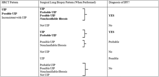
Diagnostic algorithm for idiopathic pulmonary fibrosis (IPF). Patients with suspected IPF should be carefully evaluated for identifiable causes of interstitial lung disease (ILD). In the absence of an identifiable cause for ILD, an HRCT demonstrating UIP pattern is diagnostic of IPF. In the absence of UIP pattern on HRCT, IPF can be diagnosed by the combination of specific HRCT and histopathologic pattern. The accuracy of the diagnosis of IPF increases with multidisciplinary discussion among ILD experts [5]
Table 1.1
High-resolution computed tomography criteria for UIP pattern (2011)
UIP pattern (all four features) | Possible UIP pattern (all three features) | Inconsistent with UIP pattern (any of the seven features) |
|---|---|---|
Subpleural, basal predominance | Subpleural, basal predominance | Upper or mid-lung predominance |
Reticular abnormality | Reticular abnormality | Peribronchovascular predominance |
Honeycombing with or without traction bronchiectasis | Absence of features listed as inconsistent with UIP pattern (see third column) | Extensive ground-glass abnormality (extent > reticular abnormality) |
Absence of features listed as inconsistent with UIP pattern (see third column) | Profuse micronodules (bilateral, predominantly upper lobes) | |
Discrete cysts (multiple, bilateral, away from areas of honeycombing) | ||
Diffuse mosaic attenuation/air trapping (bilateral, in three or more lobes) | ||
Consolidation in bronchopulmonary segment(s)/lobe(s) |
Table 1.2
Combination of high-resolution computed tomography and surgical lung biopsy for the diagnosis of IPF (requires multidisciplinary discussion)
 |
Stay updated, free articles. Join our Telegram channel

Full access? Get Clinical Tree


