Fig. 4.1
Scheme of autonomic innervation of the heart. The cardiac sympathetic ganglia consist of cervical ganglia, stellate ganglia, and thoracic ganglia. Parasympathetic innervation comes from the vagal nerve (Reproduced with permission from Shen et al. [12])
Atrial ganglionated plexuses were identified on (1) the superior surface of the right atrium, (2) the superior surface of the left atrium, (3) the posterior surface of the right atrium, (4) the posterior medial surface of the left atrium (the latter two fuse medially where they extend anteriorly into the interatrial septum), and (5) the inferior and lateral aspect of the posterior left atrium. Ventricular ganglionated plexuses were located in fat (1) surrounding the aortic root, (2) at the origins of the right and left coronary arteries (the latter extending to the origins of the left anterior descending and circumflex coronary arteries), (3) at the origin of the posterior descending coronary artery, (4) adjacent to the origin of the right acute marginal coronary artery, and (5) at the origin of the left obtuse marginal coronary artery [6].
Sympathetic influences on cardiac electrophysiology are complex and can be modulated by myocardial function. In the normal heart, sympathetic stimulation shortens action potential duration [4] and reduces transmural dispersion of repolarization [15]. In contrast, in pathological states such as heart failure (HF) [16] and long QT syndrome (LQTS) [17], sympathetic stimulation is a potent stimulus for the generation of arrhythmias, perhaps by enhancing the dispersion of repolarization or by generation of afterdepolarizations. In the ventricles, vagal stimulation prolongs action potential duration and effective refractory period [4, 18], whereas in the atria, vagal activation reduces the atrial effective refractory period [19, 20], augments spatial electrophysiological heterogeneity [21], and promotes early afterdepolarization (EAD) toward the end of phase 3 in the action potential [22]. This differential effect may explain why parasympathetic stimulation is proarrhythmic in the atria but antiarrhythmic in the ventricles, whereas sympathetic stimulation seems to be proarrhythmic for both chambers [23].
What happens when we apply the knowledge about the autonomic nervous system to ventricular arrhythmias?
Ischemic Cardiomyopathy
Experimentally, sympathetic stimulation induces changes in ECG repolarization and reduction of fibrillation threshold, facilitating the initiation of VF [24]. These effects are magnified in the presence of cardiac ischemia [25]. The ischemic and infarcted myocardium becomes a substrate exquisitely sensitive to arrhythmia triggers because of not only regional cellular and tissue remodeling [26] but also heterogeneity of sympathetic nervous system innervation [27].
Contributing to the heterogeneity is the phenomenon of nerve sprouting [28, 29]. By examining explanted hearts, Cao et al. [30] found that patients who had a history of ventricular arrhythmias had augmented sympathetic nerve sprouting as compared with patients with similar structural heart disease but no arrhythmias.
They also showed that there is inhomogeneous distribution of sympathetic nerve fibers in the ventricles with myocardial injury, which is characterized by regional hyperinnervation in the perivascular regions or in the periphery of injured myocardium and regional denervation in the regions with necrosis or dense fibrosis. These results suggest that there are increased sympathetic nerves after MI and that the density of these sympathetic nerves directly correlates with the occurrence of life-threatening ventricular arrhythmia.
Both infusion of nerve growth factor into the left stellate ganglia (LSG) [28] and subthreshold electric stimulation of the LSG [31] in dogs with myocardial infarction resulted in sympathetic nerve sprouting with increased ventricular fibrillation (VF) and sudden cardiac death (SCD), suggesting a causal relationship. Follow-up studies demonstrated that myocardial infarction causes the upregulation of proteins that contributed to nerve growth (nerve growth factor, growth-associated protein 43, and synaptophysin) in both the infarcted site [32] and the more upstream bilateral stellate ganglia [33]. Interestingly, sympathetic nerve sprouting itself can lead to an increased incidence of VF without concomitant cardiac ischemia.
A very original study by Liu et al. [34] found that the nerve sprouting encountered in ischemia could be induced by other stimuli, for example, a high cholesterol diet. Density of growth-associated protein 43 and tyrosine hydroxylase–positive nerves in the heart was significantly higher in high cholesterol than in control rabbits. Compared with controls, the high cholesterol rabbits had longer QTc intervals, more QTc dispersion, a longer action potential duration, and increased heterogeneity of repolarization. It was also found that rabbits given a high-cholesterol diet developed myocardial hypertrophy and cardiac sympathetic hyperinnervation without coronary artery disease along with an increased vulnerability to VF [34].
The possible explanation for this finding is based on the fact that in both human and animal studies, a high cholesterol level was associated with increased oxidative stress. Oxidative stress can cause neurodegeneration, neurite retraction, and mitochondrial dysfunction of the neurons in the central nervous system. It is therefore possible that oxidative stress causes cardiac nerve injury, which triggers the re-expression of nerve growth factor or other neurotrophic factor genes in the nonneural cells around the site of injury, leading to nerve regeneration through nerve sprouting [34].
While data from anesthetized animals suggests that sympathetic nerve activity is responsible for the development of ventricular arrhythmias, a direct temporal relationship in patients has not been established. Clinically, increased sympathetic activity, as suggested by heart rate variability analysis, was found to be in the 30 min before the onset of ventricular tachyarrhythmias [35]. With direct nerve activity recordings in a canine model of SCD, Zhou et al. [36] observed that VF and SCD were immediately preceded by spontaneous sympathetic nerve discharge from the LSG. Moreover, as the stellate ganglion nerve activity can directly be recorded with thoracotomy, the same group developed a valid technique to predict susceptibility of VT and VF in the same canine model by recording subcutaneous nerve activity. The latency from the onset of stellate ganglia activity and the subcutaneous recordings of discharges to the development of ventricular tachycardia or fibrillation was about 17 s, slightly longer than the latency of 10 s to the development of atrial arrhythmias [36]. Although increased stellate ganglion nerve activity contributes to VF and SCD in myocardial infarction, acute myocardial infarction itself can cause an increase in nerve activity and nerve density of the LSG—electroanatomic remodeling [33]. This generates a vicious cycle that can lead to more ischemia, VF, and SCD.
As the vast majority of ventricular arrhythmias in the settings of ischemia are provoked by increased sympathetic activity, early treatment with beta-blockers has been proven to reduce recurrences. Furthermore, various invasive approaches have been taken to decrease the occurrence of VT or VF.
While VT ablation has been long established treatment for organized arrhythmia in chronic ischemic cardiomyopathy, the invasive ablative approach for arrhythmias in acute ischemic events—VF or polymorphic VT—has just been recently entertained. Emerging evidence in patients with ventricular fibrillation (VF) in a variety of clinical scenarios implicates an important role for triggers originating from the distal Purkinje arborization in the initiation of this malignant arrhythmia, triggers not surprisingly sensitive to high sympathetic tone. Haissaguerre’s group was able to show on 5 patients with VF post MI that ablation of the local Purkinje network allows suppression of polymorphic VT and VF [37].
Renal denervation (RDN) is a new approach to reduce sympathetic activity. RDN reduces not only renal norepinephrine spillover by 48 % but also muscle sympathetic nerve activity by 37 %. This certainly suggests a reduction in local and central sympathetic activity after RDN by a process of modulation of efferent and afferent renal signaling. On that basis, Linz et al. elegantly showed that RDN reduced the occurrence of ventricular arrhythmias and attenuated the rise in left ventricular diastolic pressure during left ventricular ischemia created by left anterior descending artery ligation in anesthetized pigs. Therefore, RDN may protect from ventricular arrhythmias during ischemic events [38]. The same group reported their results on two cases of advanced nonischemic cardiomyopathy and incessant ventricular arrhythmias resistant to ablation and/or antiarrhythmic medication. While the patients did not have acute ischemic events, the mechanism of arrhythmia shared common features, specifically increased sympathetic tone. Renal denervation was performed successfully in these very unstable patients and led to a significant reduction in their ventricular arrhythmia load [39].
Along the same lines of neuromodulation as an alternative therapy, cardiac sympathetic denervation (CSD) has been shown to be effective in animal models and case series of patients with and without cardiomyopathy. Vaseghi et al. [40] elegantly demonstrate that left and bilateral sympathectomies, involving removal of the lower third to lower half of the stellate ganglion and T2–T4 sympathetic ganglia, have a clear benefit in the setting of VT storm and VT refractory to medical therapy in a small number of patients with cardiomyopathy. In patients with VT storm, bilateral CSD is more beneficial than left CSD. The beneficial effects of bilateral CSD extend beyond the acute postsympathectomy period, with continued freedom from ICD shocks in 48 % of patients, and a significant reduction in ICD shocks in 90 % of patients.
Conversely, studies with anesthetized dogs have shown that vagal nerve stimulation (VNS) reduces the occurrence of ventricular tachyarrhythmias after acute coronary artery occlusion. In dogs that survived an acute myocardial infarction, Vanoli et al. [41] demonstrated that VNS during the process of coronary artery occlusion reduced the occurrence of VF from 100 to 10 %.
Nonischemic Cardiomyopathy
Even though the pathophysiology of nonischemic cardiomyopathy is very different from ischemic cardiomyopathy, the provoking factors for lethal arrhythmias as well as their treatment are very similar. Because the ANS activity regulates cardiac ion channel function, it is possible that specific ANS activity might be responsible for triggering cardiac arrhythmias in congestive heart failure. Ogawa et al. [42] developed a pacing-induced CHF model in dogs and simultaneously recorded the left stellate ganglion nerve activity (SGNA), vagal nerve activity (VNA), and electrocardiography before and after pacing-induced CHF. The reduction of sympathovagal balance at night in ambulatory dogs was due to reduced sympathetic discharge rather than a net increase of vagal discharge. The tachy–brady syndrome in CHF may be triggered by intermittent short burst of SGNA that resulted in tachycardia and sinus node suppression. Simultaneous sympathovagal discharge is a cause of long paroxysmal atrial tachycardia episodes. Moreover, it was again confirmed that VA is always preceded by SGNA discharges. Importantly, when SGNA occur on the background of continuous, heightened vagal tone observed in nonischemic cardiomyopathy, the risk of VAs is increased.
While the novel neuromodulation methods used to treat arrhythmias in ischemic cardiomyopathy focused on reducing sympathetic discharges in the myocardium, the overall CHF treatment shifted attention to vagal nerve stimulation (VNS).
VNS also results in modulation of the renin–angiotensin system, reduced heart rate, modulation of inflammatory cytokines, less likelihood of spontaneous or induced ventricular arrhythmias, and reduced mortality. A single-center pilot study of eight patients with severe heart failure [seven in New York Heart Association (NYHA) class III] showed that right-sided VNS with the CardioFit system (BioControl Medical, Yehud, Israel), synchronized at 70 ms after the R wave with a duty cycle of no more than 25 %, was safe and tolerable. Sinus bradycardia was noted but was not a limiting factor since it was patient discomfort that generally prevented further increase of the stimulus intensity. There were significant and clinically important improvements in NYHA class, quality of life, and echocardiogram-derived end-systolic volume when compared with baseline values [43].
Due to encouraging positive results of the above study, three other randomized trials are ongoing (NECTAR-HF, ANTHEM-HF, and INOVATE-HF) and are assessing different outcomes in different patients. The details of the stimulation protocols are not completely clear, nor is it certain whether the protocols’ specified electrode spacing, polarity, and positioning are more likely to recruit afferent or efferent vagal fibers, whether sympathetic activity will also be suppressed, or what effect there may be on ventricular contractility. However, there is hope that this will be an important step in management of congestive heart failure and associated arrhythmias through neuromodulation mechanisms.
Inherited Arrhythmia Syndromes
In the past decade, the discovery of ventricular arrhythmias and SCD in young individuals, and the fact that they could be caused by genetic abnormalities, has defined a new subset of cardiac conditions: inherited arrhythmia syndromes. Patients with inherited arrhythmia syndromes may have a “structurally normal” heart but typically have an abnormal ECG suggesting electric abnormalities with the potential for life-threatening arrhythmias.
As in previously described conditions, the sympathetic stimulation can create a substrate for micro reentry and VF or polymorphic VT.
Long QT syndromes (LQTS)
In normal individuals, high adrenergic tone or sympathetic stimulation shortens the ventricular action potential duration and hence the QT interval. In contrast, in congenital LQTS types 1 and 2, increased adrenergic tone can prolong the QT interval. However, there is some variability in terms of the degree of response to sympathetic activation depending on the type of LQTS and, thus, the type of channel and current affected. Noda et al. [44] observed that sympathetic stimulation by infusion of epinephrine caused more prominent and prolonged effects on QT prolongation in patients with congenital LQTS type 1 (characterized by an abnormality in KCNQ1 and the I Ks current) than in type 2 (characterized by an abnormality in KCNH2 and the I Kr current). In contrast, type 3 of LQTS is characterized by an abnormality on SCN5A and Na current; therefore, it responds much less to sympathetic stimulation [44–46]. As expected, LQTS 3 manifests itself by ventricular arrhythmias triggered by increased vagal tone, similar to Brugada syndrome.
The hypothesis proposed to explain the development of LQTS-related torsades de pointes (TdP) maintains that the various mutations that underlie the syndrome amplify the transmural dispersion of repolarization (TDR) by producing a net reduction in repolarizing current. Conditions leading to a reduction in I Kr (e.g., LQT2) or augmentation of late I Na (e.g., LQT3) amplify transmural electrical heterogeneities by producing a preferential prolongation of the M cell action potential. Thus, QT interval prolongation is accompanied by a dramatic increase in TDR, which creates a vulnerable window for the development of reentry across the ventricular wall. The reduction in net repolarizing current also predisposes to the development of early afterdepolarization (EAD)-induced triggered activity in M and Purkinje cells, which provide the extrasystole that triggers TdP when it arrives during the vulnerable period [47].
CPVT
Catecholaminergic polymorphic ventricular tachycardia (CPVT) is a rare syndrome characterized by bidirectional or polymorphic ventricular arrhythmias under conditions of increased sympathetic activity in patients with structurally normal hearts. Most of the patients have mutations in the genes encoding proteins involved in calcium handling from the sarcoplasmic reticulum (ryanodine receptor or calsequestrin2 mutations) with inappropriate calcium leak that generates delayed afterdepolarizations (DAD), triggered activity, and ventricular arrhythmias [48].
Recently, research groups have identified the therapeutic effects of flecainide in CPVT. Flecainide directly inhibits the ryanodine channel and suppresses DADs and triggered activity in mutant cardiomyocytes in which Ryr2 or Casq2 loci are modified. In a recent study, Watanabe et al. found that flecainide suppressed ventricular arrhythmias during exercise testing in patients with genotype-negative CPVT, similar to that in patients with genotype-positive CPVT. Flecainide was highly effective in preventing arrhythmia events during a long-term follow-up [49].
Since both LQTS and CPVT are sympathetically mediated, in order to prevent cardiac events, β-blocker pharmacotherapy is the cornerstone of medical therapy while high-risk patients will benefit from ICDs. Additionally, the left cardiac sympathetic denervation (LCSD) procedure, in which the first 3–4 thoracic ganglia are removed, has emerged as a treatment modality for patients with LQTS, and more recently patients with CPVT, who continue to have cardiac events despite maximally tolerated β-blocker therapy, and has been proved to be a safe and effective treatment option [50]. Potential adverse effects of this procedure could include Horner’s syndrome and altered sweating patterns in the face and upper extremities.
Brugada Syndrome
The Brugada syndrome is an inherited arrhythmia disorder characterized by its typical ECG changes (right bundle branch block and persistent ST segment elevation) and an increased risk of VF and SCD in young patients.
Most episodes of VF in patients with Brugada syndrome are observed during periods of high vagal tone, such as at rest, during sleep, or from 12 am to 6 am. Similarly to LQTS 3, sudden increase of vagal activity just before the episodes of VF has been noted. This suggests that an increased vagal tone or decreased sympathetic tone may be important mechanisms in the arrhythmogenesis of this lethal disease [51]. Additionally, as the epicardium of right ventricular outflow tract serves as a substrate that mediates and sustains ventricular arrhythmias triggered by altered autonomic tones, the arrhythmia may be ablated successfully while also eliminating the repolarization abnormalities [52].
Arrhythmogenic right ventricular cardiomyopathy (ARVC)
is a frequent underlying disease in young patients with ventricular tachycardia (VT) and sudden death. These arrhythmias often occur during physical exercise or mental stress and may be provoked by intravenous catecholamine infusion during electrophysiological study (catecholamine sensitivity). In contrast, VAs are frequently suppressed by an antiarrhythmic drug regimen with antiadrenergic properties [53].
Wichter et al. [53] have investigated the neuronal reuptake of norepinephrine and beta-adrenergic receptor density in eight patients with ARVC using quantitative [11] PET C-HED and [11] C-CGP-12177 as compared to twenty-nine matched controls.
The results provide clear evidence of abnormal sympathetic myocardial innervation in patients with ARVC and demonstrate a severe and highly significant reduction of postsynaptic beta-adrenergic receptor density. Additionally, this confirms regional reduction of transporter-mediated neuronal catecholamine reuptake in ARVC. Moreover, increased synaptic concentrations of norepinephrine not only may increase the risk of ventricular arrhythmias but also may contribute to progression of myocardial atrophy mediated by apoptotic cell death.
While ablation of reentrant arrhythmias is performed in highly experienced centers by accessing the epicardial surface of the scarred right ventricle [54], implantable defibrillators and treatment with beta-blockers and class III antiarrhythmic agents remain the mainstay of management at this time.
Idiopathic VF with repolarization abnormalities,
the so-called J-point elevation syndromes, is characterized by ST segment elevation in the inferior and/or lateral leads. While this ECG finding is fairly frequently seen in young athletic adults, identifying the high-risk population is, of course, extremely important as those are at risk for sudden cardiac death due to VF. While most of the congenital abnormalities bear high risk of arrhythmias in the settings of high adrenergic tone, this rare syndrome seems to be triggered by increased vagal tone while exercise and increased sympathetic tone or isoproterenol infusion decrease the chance of arrhythmias.
For example, Mizumaki et al. [55] recently observed that in patients with idiopathic VF as compared with control subjects, J-wave augmentation was associated with an increase in vagal activity. This data suggests a critical role of cardiac ANS in the occurrence of VF in patients with J-wave syndromes. The differential responses of characteristic ECG changes to autonomic input may provide a useful tool in the identification of high-risk patients within the broad population of healthy individuals with this specific ECG pattern. While at this time autonomic modulation approaches for this condition are not available, treatment options are on the horizon.
Premature ventricular contractions
can, as mentioned above, also pose diagnosis and management problems, as their impact on patients varies from asymptomatic PVCs to mild palpitations to acquired “PVC-induced cardiomyopathy” with heart failure and even VF and sudden cardiac death.
In the setting of ischemic and nonischemic cardiomyopathy, the PVCs seem to originate from areas around the scar and surviving Purkinje fibers. Their triggering factors and treatment options, including neuromodulation techniques, are similar to the management of VF and VT.
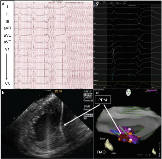
When conservative medical therapy fails, ablation of these PVCs provides additional and significant benefit. Marchlinski et al. recently published a retrospective study focused on the location of the PVCs which induce VF both structurally normal and abnormal hearts.
Ventricular fibrillation and polymorphic ventricular tachycardia (PMVT) triggered by monomorphic PVCs were the presenting arrhythmias in 30 of 1132 patients (2.7 %) undergoing ablation for VT/PVCs. Analysis of the study cohort revealed that in 21 patients, VF/PMVT occurred in the setting of ischemic (n = 14) or nonischemic CMP (n = 7). In 9 patients, there were no cardiac structural abnormalities, and VF/PMVT was idiopathic. The PVC triggers were found to originate from the Purkinje network (n = 9), left and right ventricle papillary muscles (n = 8), left ventricular outflow tract (LVOT) (n = 9), and other low-voltage areas (without evidence of Purkinje system involvement [scar triggers]; n = 4). Each distinct anatomic area of origin was associated with VF/PMVT trigger in patients with and without heart disease. Acute PVC elimination was accomplished in 26 patients (87 %) with a significant decrease in PVCs in another 3 patients (97 %). In structurally normal heart patients, the PVCs originated predominantly from left ventricular outflow tract as well as Purkinje fibers. Conversely, in patients with cardiomyopathies, the VF inducing PVCs originated from scar borders and Purkinje fibers as expected. While no cases of VF triggered by PVCs from the papillary muscles (PM) have been previously described, this group found six patients with cardiomyopathy and two patients with structurally normal heart and reproducible arrhythmias originate in the PM (Figs. 4.2 and 4.3) [56].