Abstract
Background
Coronary angiography (CA) has been the gold standard technique for studying coronary artery disease. It is based on the analysis of bidimensional orthogonal projections that may not be optimal to estimate determinate coronary segments. Rotational angiography “Xperswing” (DARCA) is a new technique that allows the visualization of the coronary arteries from multiple views, with a single contrast injection. The aim of this study is to evaluate the coronary lesions quantification with DARCA.
Methods
Quantitative coronary analysis of significant coronary stenosis (> 50%) was performed. Every lesion was measured in two different projections: the “optimal projection”, obtained by DARCA and defined by the operator as the one with a better lesion qualification, and the “standard projection”, corresponding to the usual projection closer to the optimal one in obliquity and angulation. Measures were performed twice and by two independent operators. Intra- and inter-observer correlation was estimated by Kappa index and variables were compared with t Student test (SPSS 14.0).
Results
205 lesions in 147 patients were analyzed. Kappa coefficient intra-observer was 0.80 and 0.86 respectively with an inter-observer correlation index of 0.72. Lesion length and maximal diameter of the vessel were significantly greater in the group of RA. In the segments analysis, calculated length was longer for the first diagonal branch, first marginal obtuse artery, middle circumflex, middle and distal RCA and posterior descending artery, with greater reference diameters for proximal LAD and distal RCA. There were no significant differences for coronary stenosis grade.
Conclusions
RA XperSwing provides a better visualization of coronary arteries improving lesions characterization, with longer measured lesions length and greater vessel diameters, especially in coronary segments with more angulation.
1
Background
Coronary angiography (CA) remains as the gold standard technique for the diagnosis and treatment of coronary lesions. It is carried out in multiple predefined bidimensional stationary views, at different angulations around the patient, and provides only a limited number of projections of the vascular tree despite the use of a considerable amount of contrast medium and radiation exposure. Over the years, studies of angioscopy and pathologic analysis have demonstrated the limited diagnostic accuracy of conventional angiography, especially in overlapped vessels and angulated segments .
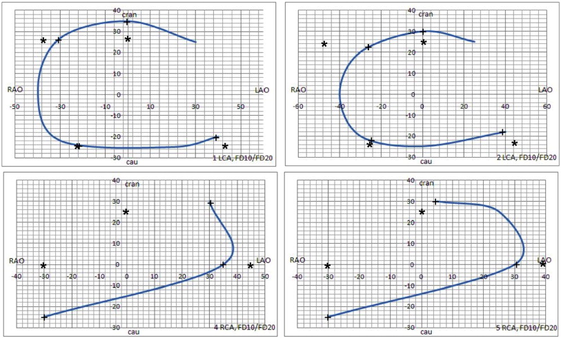
Dual-axis rotational coronary angiography (DARCA) XperSwing® is an image acquisition technique in which the X-ray system swings around the patient mixing rotation and angulation during the acquisition of a single run. DARCA is available for systems with monoplane detector Allura Xper FD10 o FD 20 (Philips Medical Systems, The Netherlands) and provides different trajectories for the left and right coronary arteries using a rapid isocentric rotation of the imaging camera. The rotational representation of the coronary tree allows a better three-dimensional understanding of the spatial relationships of coronary tree branches compared to the traditional CA . XperSwing contains 7 different trajectories for equipment FD 10 and 6 for equipment FD 20 ( Fig. 1 and Table 1 ). With this system, the complete study may be performed with a total contrast amount of 35 ml, acquiring 149 images (87 for left coronary artery and 62 for right coronary artery) with an exposure time of 9.9 s (considering exclusively the acquisition phase) ( Fig. 2 ).
| Optimal projection | Standard projection | P | |
|---|---|---|---|
| Global measures | |||
| Minimum lumen diameter (mm) | 1.13 ± 0.39 | 1.17 ± 0.47 | 0.33 |
| Lesion length (mm) | 9.74 ± 2.55 | 7.83 ± 2.81 | 0.01 |
| Maximal lumen diameter (mm) | 3.39 ± 0.49 | 3.21 ± 0.58 | 0.00 |
| % Diameter | 50.97 ± 10.70 | 49.05 ± 11.58 | 0.11 |
| % Stenosis | 82.29 ± 9.07 | 81.58 ± 7.67 | 0.46 |
| Left main (6) | |||
| Length | 9.21 ± 1.71 | 8.85 ± 1.95 | 0.69 |
| Maximal lumen diameter | 4.34 ± 0.58 | 4.16 ± 0.37 | 0.18 |
| Minimum lumen diameter | 1.76 ± 0.41 | 1.61 ± 0.51 | 0.87 |
| % Diameter | 42.34 ± 8.93 | 41.92 ± 9.75 | 0.94 |
| % Stenosis | 66.54 ± 8.49 | 65.87 ± 8.58 | 0.89 |
| Proximal LAD (26) | |||
| Length | 9.09 ± 2.53 | 8.93 ± 2.38 | 0.13 |
| Maximal lumen diameter | 3.54 ± 0.31 | 3.11 ± 0.60 | 0.01 |
| Minimum lumen diameter | 1.02 ± 0.74 | 1.20 ± 0.62 | 0.78 |
| % Diameter | 62.50 ± 7.20 | 63.28 ± 8.93 | 0.69 |
| % Stenosis | 84.92 ± 8.28 | 86.49 ± 7.39 | 0.39 |
| Middle LAD (29) | |||
| Length | 9.54 ± 2.28 | 8.72 ± 3.66 | 0.06 |
| Maximal lumen diameter | 3.55 ± 0.42 | 3.56 ± 0.44 | 0.61 |
| Minimum lumen diameter | 1.06 ± 0.29 | 1.15 ± 0.33 | 0.87 |
| % Diameter | 53.87 ± 8.39 | 54.31 ± 5.90 | 0.57 |
| % Stenosis | 86.82 ± 7.49 | 86.97 ± 6.79 | 0.73 |
| Distal LAD (16) | |||
| Length | 8.02 ± 0.49 | 6.98 ± 1.60 | 0.15 |
| Maximal lumen diameter | 3.17 ± 0.48 | 2.96 ± 0.14 | 0.12 |
| Minimum lumen diameter | 0.86 ± 0.31 | 0.97 ± 0.65 | 0.49 |
| % Diameter | 59.38 ± 6.10 | 61.38 ± 7.39 | 0.38 |
| % Stenosis | 95.47 ± 5.29 | 98.69 ± 6.59 | 0.43 |
| First diagonal branch (8) | |||
| Length | 10.02 ± 0.56 | 7.98 ± 1.60 | 0.00 |
| Maximal lumen diameter | 2.88 ± 0.23 | 2.76 ± 0.48 | 0.21 |
| Minimum lumen diameter | 0.79 ± 0.36 | 0.68 ± 0.38 | 0.44 |
| % Diameter | 57.51 ± 5.31 | 56.89 ± 6.92 | 0.51 |
| %Stenosis | 91.64 | 90.04 | 0.19 |
| Proximal Cx (23) | |||
| Length | 9.43 ± 2.52 | 9.19 ± 2.67 | 0.74 |
| Maximal lumen diameter | 4.02 ± 0.41 | 4.12 ± 0.28 | 0.81 |
| Minimum lumen diameter | 1.87 ± 0.59 | 1.59 ± 0.49 | 0.23 |
| % Diameter | 45.21 ± 5.27 | 47.58 ± 3.89 | 0.68 |
| % Stenosis | 72.35 ± 6.29 | 75.56 ± 7.36 | 0.42 |
| Middle Cx (20) | |||
| Length | 9.79 ± 1.64 | 6.08 ± 0.56 | 0.00 |
| Maximal lumen diameter | 3.39 ± 0.22 | 3.24 ± 0.44 | 0.19 |
| Minimum lumen diameter | 1.49 ± 0.32 | 1.59 ± 0.39 | 0.32 |
| % Diameter | 52.29 ± 4.58 | 61.29 ± 5.69 | 0.11 |
| % Stenosis | 85.21 ± 5.29 | 98.07 ± 9.28 | 0.09 |
| Distal Cx (8) | |||
| Length | 9.68 ± 1.51 | 8.64 ± 1.28 | 0.08 |
| Maximal lumen diameter | 3.16 ± 0.44 | 2.99 ± 0.32 | 0.43 |
| Minimum lumen diameter | 1.04 ± 0.38 | 0.98 ± 0.59 | 0.49 |
| % Diameter | 51.24 ± 5.39 | 53.29 ± 8.39 | 0.39 |
| % Stenosis | 82.29 ± 3.30 | 85.69 ± 6.12 | 0.65 |
| Marginal obtuse branch (12) | |||
| Length | 11.02 ± 2.52 | 8.19 ± 2.67 | 0.00 |
| Maximal lumen diameter | 2.98 ± 0.43 | 2.91 ± 0.29 | 0.81 |
| Minimum lumen diameter | 0.99 ± 0.29 | 1.13 ± 0.48 | 0.66 |
| % Diameter | 50.98 ± 5.38 | 51.49 ± 5.99 | 0.41 |
| %Stenosis | 83.29 ± 7.29 | 84.98 ± 6.39 | 0.34 |
| Proximal RCA (8) | |||
| Length | 9.22 ± 1.79 | 8.65 ± 1.35 | 0.06 |
| Maximal lumen diameter | 3.97 ± 0.46 | 3.70 ± 0.29 | 0.11 |
| Minimum lumen diameter | 1.57 ± 0.39 | 1.63 ± 0.38 | 0.73 |
| % Diameter | 47.39 ± 3.73 | 49.82 ± 4.38 | 0.38 |
| % Stenosis | 75.57 ± 8.25 | 78.78 ± 7.36 | 0.44 |
| Middle RCA (21) | |||
| Length | 10.93 ± 2.96 | 7.65 ± 2.37 | 0.00 |
| Maximal lumen diameter | 3.67 ± 0.33 | 3.56 ± 0.42 | 0.11 |
| Minimum lumen diameter | 1.37 ± 0.48 | 1.43 ± 0.29 | 0.62 |
| % Diameter | 49.58 ± 4.48 | 51.32 ± 2.49 | 0.45 |
| % Stenosis | 79.71 ± 3.93 | 82.51 ± 3.12 | 0.21 |
| Distal RCA (12) | |||
| Length | 9.89 ± 2.15 | 8.73 ± 2.52 | 0.01 |
| Maximal lumen diameter | 2.87 ± 0.37 | 2.52 ± 0.29 | 0.03 |
| Minimum lumen diameter | 1.12 ± 0.39 | 1.19 ± 0.48 | 0.67 |
| % Diameter | 49.87 ± 4.19 | 51.21 ± 5.60 | 0.30 |
| % Stenosis | 80.18 ± 7.12 | 82.336.89 | 0.65 |
| Posterolateral artery (6) | |||
| Length | 8.89 ± 1.98 | 7.21 ± 1.46 | 0.06 |
| Maximal lumen diameter | 2.77 ± 0.37 | 2.62 ± 0.29 | 0.45 |
| Minimum lumen diameter | 0.78 ± 0.28 | 0.93 ± 0.16 | 0.79 |
| % Diameter | 59.21 ± 2.15 | 62.38 ± 7.39 | 0.41 |
| % Stenosis | 95.02 ± 5.49 | 99.03 ± 6.59 | 0.38 |
| Posterior descending artery (10) | |||
| Length | 10.85 ± 2.53 | 8.84 ± 2.27 | 0.01 |
| Maximal lumen diameter | 2.69 ± 0.27 | 2.67 ± 0.26 | 0.82 |
| Minimum lumen diameter | 0.89 ± 0.31 | 0.72 ± 0.59 | 0.57 |
| % Diameter | 50.31 ± 5.46 | 49.29 ± 6.99 | 0.48 |
| % Stenosis | 80.89 ± 7.89 | 79.37 ± 6.25 | 0.41 |
Stay updated, free articles. Join our Telegram channel

Full access? Get Clinical Tree


