Fig. 7.1
ECG demonstrating sinus rhythm with lateral T wave inversion and ventricular ectopy
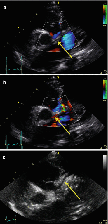
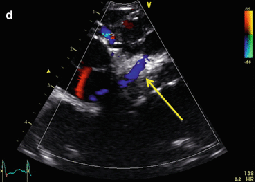
Fig. 7.2
Initial echocardiogram demonstrating apparently normally arising LCA (arrow) from the aorta (a) with antegrade flow (b). Subsequent echocardiogram demonstrating true left coronary artery (c) and retrograde flow (d)
A diagnosis of dilated cardiomyopathy was made. The child was intubated and ventilated, and managed with inodilation, adrenaline and isoprenaline for bradycardia. A cardiomyopathy screen returned normal. The child’s condition did not improve and the ECG developed progressive ST changes consistent with ischaemia as well as frequent ventricular ectopics and runs of tachycardia. The echocardiogram was repeated specifically to re-assess the LCA. The original appearance of a normal connection and direction of flow could be reproduced but was in fact misleading. The vessel originally thought to be the LCA was instead a small branch of the right coronary artery (RCA) coursing in the direction the LCA would ordinarily take, and therefore showed antegrade flow. A much larger vessel, the LCA, could be seen arising from the pulmonary artery. The direction of flow was retrograde (see Fig. 7.2c, d). A diagnosis of anomalous left coronary artery from the pulmonary artery (ALCAPA) was made. Reduction of the adrenaline and isoprenaline infusions resulted in less ventricular ectopy, and surgery was undertaken the same day.
At operation there was extensive collateralisation around the aorta and pulmonary artery, possibly accounting for the initial echocardiographic impression of a normal LCA. The LCA was transferred to the aorta, but despite good flow being seen on epicardial echocardiogram myocardial performance remained poor. The child was weaned off cardiopulmonary bypass, but due to the advanced state of left ventricular damage suffered persistent ventricular fibrillation and was placed on extra-corporeal membrane oxygenation (ECMO) prior to transfer to the intensive care unit. A week later the patient was separated from ECMO; however, she remained fully dependent on inotropic support and positive pressure ventilation, and experienced frequent ventricular tachyarrhythmias requiring resuscitation. Five weeks post-operatively one such arrest necessitated re-institution of EMCO support. Ventricular dysrhythmias continued despite a continuous amiodarone infusion and fastidious management of electrolyte balance.
There were grave concerns over the extent of irreversible myocardial damage by the time of presentation, and that transplantation represented the only possibility of survival. The patient was transferred to a transplant centre and support converted to a biventricular Berlin Heart. She was listed for urgent cardiac transplantation; however, she developed complications of mechanical circulatory support, including bacterial infection and evidence of neurologic injury manifested as choreoathetoid movements upon weaning of sedation. She also had persistent atelectasis of the left lower lobe due to gross cardiomegaly-induced compression of the left main bronchus. An initial CT demonstrated only a small sub-acute subdural haemorrhage; however, she subsequently became less responsive and developed fixed dilation of the left pupil. A repeat CT demonstrated a devastating intracranial haemorrhage. Following discussion with the patient’s parents, active support was withdrawn and care re-orientated.
Discussion
Rapid diagnosis of ALCAPA is important for two reasons. Firstly, severe, irreversible damage to left ventricular myocardium may occur, resulting in death in up to 80 % of patients by 1 year of life. Secondly, this outcome can be avoided by prompt re-implantation of the left coronary artery into the aorta.
Normal Anatomy and Physiology
The LCA ordinarily divides into the left anterior descending (LAD) and the circumflex (Cx) arteries. The LAD artery lies anteriorly in the interventricular groove, while the circumflex artery courses posteriorly between the left atrium and ventricle. They account for the coronary arterial blood supply to the left atrium and ventricle. These arteries lie within the epicardium and give rise to smaller branches that penetrate the myocardium. The contraction of the myocardium in systole results in compression of these smaller branches, the highest degree of compression being in the subendocardial regions. Consequently, the filling of the coronary arteries is restricted during cardiac systole. In fact, filling of the LCA occurs exclusively in diastole. Subsequently, the diastolic aortic pressure and duration of diastole are important determinants of coronary artery perfusion. Any significant fall in the diastolic pressure may lead to ischaemia, the subendocardium being the area most susceptible to injury.
Pathophysiology
The exact location of the LCA ostium in ALCAPA varies. The most common site is the posterior-facing sinus of the pulmonary artery. Sometimes the LAD or Cx arises alone. Ectopic location is also possible with any coronary artery, with origin from the anterior left non-facing sinus, the main pulmonary artery, or the pulmonary branches reported.
< div class='tao-gold-member'>
Only gold members can continue reading. Log In or Register to continue
Stay updated, free articles. Join our Telegram channel

Full access? Get Clinical Tree


