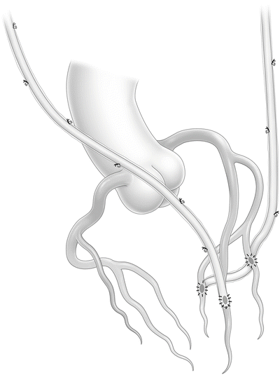
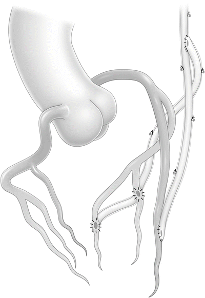
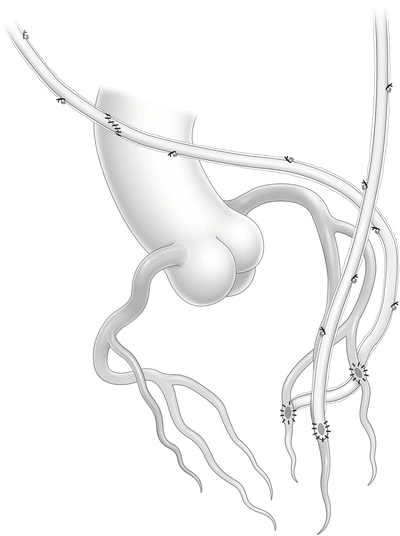
Fig. 13.1
Left internal thoracic artery is grafted to the left anterior descending artery. The right internal thoracic artery is grafted to the circumflex artery
We usually use in situ LITA to LAD because of its straight course parallel to the LAD. An in situ LITA has adequate length to reach any part of the LAD. However, because the distal part of the ITA is thin and its media predominantly muscular, use of the distal part of the LITA should be avoided. When the LAD has a long, diffusely diseased lesion, the in situ LITA can be used to perform long-segmental reconstruction of the LAD with onlay patch grafting [22]. The in situ RITA does not, however, have sufficient length to reach the distal LAD for long-segmental reconstruction. Another option for in situ LITA to LAD is that it is possible to perform sequential grafting to the diagonal branch. When the diagonal branch lies adjacent to the LAD, the LITA goes almost straight to the LAD. When the angle between the diagonal branch and the LAD is wide, sequential grafting is not recommended because the LITA may be bent and thus has a risk of kinking.
When the in situ RITA is anastomosed to the LCX territory, it is passed through the transverse sinus. When side branches of the RITA are divided, a metal clip should not be used because the clip may come off when the graft is passed through the transverse sinus and lead to bleeding behind the aorta. It is preferable to harvest the ITA by skeletonizing it with an ultrasonic scalpel. The RITA is completely mobilized from the level of the subclavian vein, and the right internal thoracic vein is divided before its connection to the brachiocephalic vein. The pericardium of the right upper portion is incised anterior to the right phrenic nerve. The distal anastomosis of in situ RITA to the proximal part of the LCX with off-pump technique can be technically demanding. We always apply a commercially available heart positioner and stabilizer to the heart and often widely open the right pleura to avoid right heart compression and obstruction of venous return. Anastomosis is performed with an 8–0 polypropylene running suture using parachute technique. This configuration is much safer than in situ RITA to LAD at reoperation.
The in situ RITA is occasionally used for a high lateral or diagonal branch running in front of the aorta. With this strategy, the in situ RITA should be protected with the thymus at the end of the procedure in order to prevent damage at reoperation.
13.3.2 In Situ RITA to LAD and In Situ LITA to LCX (Fig. 13.2)

Fig. 13.2
Right internal thoracic artery is grafted to the left anterior descending artery. The left internal thoracic artery is grafted to the circumflex artery
When the in situ RITA is anastomosed to the LAD, it is directed anterior to the aorta. With this strategy, the length of the in situ RITA should be checked to ensure that it has sufficient length, which it does in nearly all patients, to reach the LAD. In some cases, however, increased volume of the right heart can increase the distance between the LAD and the root of the RITA. Patients with right heart failure can be improved after complete revascularization. Pevni et al. suggested that in situ RITA to LAD not be performed in patients with a short RITA, a very long ascending aorta, an enlarged right ventricle, or an LAD anastomotic site that is too distal or unpredictable or in patients with high probability of repeat surgery, such as valve operations [23].
This strategy is especially useful in patients with unstable left main or bifurcation disease using off-pump technique [24]. The LAD is first revascularized using the in situ RITA with minimal rotation of the heart. After completing LAD revascularization, the LCX can be revascularized using the in situ LITA for hemodynamic stability. The patency rate of the in situ RITA is comparable to that of the in situ LITA when anastomosed to the LAD [25]. Furthermore, in our analysis, the patency rate of in situ RITA to LAD was superior to that of in situ RITA to LCX [26].
In situ RITA grafts are sometimes at risk of injury at reoperation. The RITA should be wrapped in thymic tissue and covered with mediastinal fat at the end of the surgery to prevent injury upon reopening [27]. If adhesions between the in situ RITA and sternum are severe at reoperation, establishment of cardiopulmonary bypass with femoral or axillary cannulation is recommended to decompress the heart.
When in situ LITA is used for the LCX, multiple sequential anastomoses can be performed. If the LITA is extremely long, it is possible to anastomose it to the posterolateral branch of the RCA. When sequential anastomoses are carried out with the in situ LITA, the first anastomosis should be done between the most proximal branch of the LCX and the LITA in side-to-side fashion because the length and course of the LITA are easily chosen. We prefer creating a perpendicular anastomosis in a diamond shape to a parallel anastomosis. The second or third anastomosis is made sequentially to the distal branches. The terminal anastomosis is done either perpendicularly or parallel at the discretion of the surgeon.
13.4 Composite Grafting with BITAs
When the in situ RITA cannot reach the LAD or LCX, the RITA is frequently used as a free graft. When the LCX has multiple lesions, sequential grafting is performed using a free RITA graft. With this strategy, proximal anastomosis of the free RITA is performed to the in situ LITA, to another graft (RA or SVG), or directly to the aorta. In contrast, the LITA is used as a free graft in a limited number of situations, for example, where there is injury during harvesting, subclavian artery stenosis, or radiation injury.
Free RITA-to-aorta anastomosis is rarely performed because the patency rate of the free RITA proximally anastomosed to the aorta may be inferior to that of the LITA anastomosed to the aorta. The poor graft patency of the free RITA anastomosed to the aorta is considered to be due to mismatch between the aorta and the conduit wall and a difference in the flow pattern [8].
Because BITA for the left coronary system has shown superior long-term survival compared with single ITA, a free ITA should be used for left coronary artery. Making a Y- or T-shaped anastomosis between the free ITA and the in situ ITA is performed either before or after construction of the distal anastomoses. We prefer creating a Y-shaped anastomosis after performing all distal anastomoses because at that point decisions regarding the length and course of the free ITA graft are more easily made. A benefit of this configuration is that fewer grafts are required.
Multiple sequential distal anastomoses between the free ITA and branches of the LCX with off-pump technique are considered to be a more complex procedure because the free ITA is smaller and shorter than other grafts. However, the ITA is rarely atherosclerotic and is easy to handle when taken with skeletonization. Tatoulis et al. demonstrated that the 10-year patency rates of in situ (n = 450, 89 %) and free (n = 541, 91 %) RITA grafts were similar (p = 0.44) [28]. Furthermore, in our previous analysis, follow-up angiographic results of sequential grafts were as good as those of individual grafts [29]. Competitive or reverse flow due to a lack of stenosis in a native coronary artery could be the potential cause of occlusion in composite grafts [30].
13.4.1 In Situ LITA to LAD and Free RITA to LCX (Fig. 13.3)

Fig. 13.3
Left internal thoracic artery is grafted to the left anterior descending artery. The free right internal thoracic artery or radial artery is sutured to the in situ left internal thoracic artery as a Y-graft to multiple circumflex branches
We typically use the RITA as free graft because multiple sequential grafts to lateral or posterior vessels can be performed with this graft. A metal cannula is inserted into the proximal end of the free RITA and the vessel ligated, and milrinone solution is injected to dilate the graft. One side of the graft is marked with crystal violet to prevent twisting. When sequential distal anastomoses are carried out with the free RITA, the first anastomosis is performed side to side to the most proximal branch in diamond-shaped fashion. The second or third anastomosis is created sequentially to the distal branches. The terminal anastomosis is done either perpendicular or parallel. The free RITA can reach the posterior descending artery when its proximal anastomosis is carried out on the left ITA [31].
The most popular site of proximal anastomosis of the free RITA is the in situ LITA using a T or Y configuration, depending on the length of the free RITA graft. Tector et al. demonstrated excellent results using BITA T grafts [9], reporting that sewing the BITAs at right angles (T shaped) preserves valuable proximal length and prevents kinking. The T anastomosis can be located proximally or distally to accommodate the position of the first distal coronary anastomosis. We prefer a Y-shaped anastomosis because of the smaller angle between the in situ LITA and the free RITA, which may lead to smoother blood flow. The best anastomotic site is at the level of the pulmonary artery. Calafiore et al. showed that Y grafting using BITAs had the same angiographic and clinical results as in situ BITAs [32]. Anastomosis between two ITAs is not as difficult because the wall characteristics are the same. We use an 8–0 polypropylene running suture for this anastomosis.
13.4.2 In Situ RITA to LAD and Free LITA to LCX
Based on the same anatomical characterization of the BITAs, composite grafting with in situ RITA and free LITA may be possible [23]. However, this configuration is extremely rare and is limited to occasions when the LITA is not available for in situ grafting because of injury during harvesting, subclavian artery stenosis, or radiation injury due to a mediastinal tumor or breast cancer. Making a T or Y anastomosis between the in situ RITA and the free LITA is performed at the anterior surface of the heart and near to the distal anastomosis of the in situ RITA. Care should be taken not to kink or bend the in situ RITA if the free LITA is short.
13.5 ITA and RA Composite Grafting
The RA is the second or third most frequently used arterial graft. It is a muscular artery and usually has a thick media comprising mostly smooth muscle cells. Thus, it sometimes has an atherosclerotic intima and macroscopic calcification. Because it always needs antispasmodic preparation, we inject milrinone solution directly into the RA and administer diltiazem systemically. Recently, the distal side of the RA has sometimes been used for catheterization in patients undergoing coronary angiography or coronary intervention, so care must be taken not to use distal part aspect for revascularization in these patients.
The RA is easily handled, and when it is skeletonized for harvest, sequential anastomoses for two or three distal coronary arteries can easily be performed. Various sites of proximal anastomosis of the RA have been debated. Some prefer anastomosing the RA directly to the aorta, and others prefer a T or Y ITA composite. In some cases, a straight graft (I-composite graft) with the in situ RITA is possible. If multiple distal anastomoses using the RA are necessary, composite grafting should be considered because it may not reach the aorta. Several authors have demonstrated favorable clinical and angiographic results using the RA [33]. Distal runoff, competitive flow, and proximal anastomosis site are considered to influence the patency of the RA. In our analysis, non-ITA graft, mild target stenosis, and multiple grafts from a single inflow source were independent predictors of graft deterioration [30]. In other words, the RA graft, when it is used in composite and multiple grafts from a single inflow source, tends to deteriorate in targets in which stenosis is not severe. We have recently begun to limit the use of RA to LCX with severe stenosis in non-elderly patients.
13.5.1 In Situ LITA to LAD and RA to Circumflex Artery (Fig. 13.3)
Kobayashi and Nakajima have had extraordinary experiences of in situ LITA and RA composite grafting [34, 35]. The RA is skeletonized and harvested. The length of the RA is almost always greater than 20 cm and is as easily handled as a skeletonized SVG, making the creation of multiple RA grafts easier than free RITA grafting.
When the RA used as a T- or Y-composite graft with the in situ LITA, distal anastomosis is performed first. When sequential anastomoses are carried out with the RA, the first anastomosis should be done to the most proximal branch in side-to-side, diamond-shaped fashion, followed by a second or third anastomosis to the distal branches created sequentially. The terminal anastomosis is created either perpendicular or parallel. Because the wall of the RA is thicker than that of other vessels, care should be taken to pass the needle from intima to adventitia, especially in side-to-side anastomosis. The RA is long enough to reach the posterior descending artery when its proximal anastomosis is to the LITA. Although proximal anastomosis of the RA to the in situ LITA is not difficult, it should be performed carefully because there is a size discrepancy between the ITA and RA. The length of the ITA incision should be sufficiently matched to the RA (sometimes requiring greater than 5 mm) so as not to create stenosis of the ITA just distal to the RA-ITA anastomosis. The site of the T or Y anastomosis should be located according to the position of the first distal coronary anastomosis.
13.5.2 In Situ RITA Combined with RA to Circumflex Artery (Fig. 13.4)

Fig. 13.4
Left internal thoracic artery is grafted to the left anterior descending artery. The in situ right internal thoracic artery and radial artery are anastomosed as an I-graft to multiple circumflex branches
Some surgeons prefer an I-composite to a Y-composite in situ RITA-to-RA graft [36]. This configuration has several advantages. First, in situ LITA to LAD is secured and not traumatized by any other grafts that may cause kinking or twisting. In addition, I-composite grafts are long enough to reach all coronary territories. Only the upper one-third of the entire length of the RITA is used; the rest of the RITA is maintained to preserve the blood supply to the body of the sternum on the right side. The RITA and RA are anastomosed in end-to-end or end-to-side fashion. An end-to-side anastomosis consisting of the distal end of the RITA and the side of the RA can prevent a purse-string effect at the anastomosis and is thus our preference. The I-composite graft can reach posterolateral and inferior branches simultaneously; however, the route of the graft (clockwise or counterclockwise orientation) must be determined. According to Kobayashi et al. [37], the route of the I-composite graft should be determined on the basis of the degree of stenosis of the terminal coronary artery anastomosed to the end of the conduit; the terminal coronary artery should have severe stenosis to avoid competitive reverse flow. The angiographic patency rate of I-composite grafts has been shown to be 99.2 % early postoperatively [37]. When an I-composite graft runs anterior to the heart, it should be wrapped in thymic tissue and covered with mediastinal fat at the end of the surgery to prevent injury in the event of reopening.
Stay updated, free articles. Join our Telegram channel

Full access? Get Clinical Tree


