Control of Ventilation and Respiratory Muscles

Chapter 6 Control of Ventilation and Respiratory Muscles


The respiratory muscles are the only muscles, along with the heart, that must work continuously, although intermittently, to sustain life. They have to repetitively move a rather complex elastic structure, the thorax, to achieve the entry of air into the lungs and thence effect gas exchange. The presence of multiple muscle groups in this system mandates that these muscles interact properly to perform their task despite their differences in anatomic location, geometric orientation, and motor innervation. They also should be able to adapt to a variety of working conditions and respond to many different chemical and neural stimuli.


This chapter describes some aspects of respiratory muscle function that are relevant to current understanding of the way these muscles accomplish the action of breathing and how their function is controlled by the respiratory centers located in the central nervous system.


The Respiratory Centers


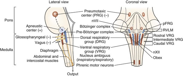
Early studies of the neural control of breathing involved the section and ablation of various brain stem structures. From these studies emerged the classical description of the neural control of breathing that required centers in the medulla for the rhythmic generation of ventilatory drive plus additional areas in the pons (traditionally known as the pneumotaxic and apneustic centers) that modulated and regulated the basic rhythm. Nowadays, the very complex and inadequately explored and understood respiratory center structure and function can be summarized as follows (Figure 6-1):


Primary centers responsible for the generation of respiratory rhythm are located in the medulla. Within the medulla, there are two bilateral aggregations of neurons having respiratory related activity.


An area of the ventrolateral medulla next to the nucleus ambiguus, the pre-Bötzinger complex, is hypothesized to be a critical site for respiratory rhythmogenesis. Current theory proposes that a group of pacemaker neurons depolarize, fire, and repolarize in a rhythmic fashion. This endogenous oscillatory activity can be modulated by afferent inputs, generating an efferent output that is translated into the respiratory drive. Apart from the pre-Bötzinger complex principally involved in controlling inspiratory motor activity, the retrotrapezoid-parafacial respiratory group (RTN/pFRG) appears to play at least a modulatory role and may be a conditional oscillator that controls active expiration.


An additional mechanism is voluntary control of the respiratory muscles, signals for which originate in the motor cortex and pass directly to the spinal motor neurons by way of the corticospinal tracts. The medullary respiratory control center is bypassed. The voluntary control competes with automatic control at the level of the spinal motor neuron.


Afferent Inputs to the Respiratory Centers


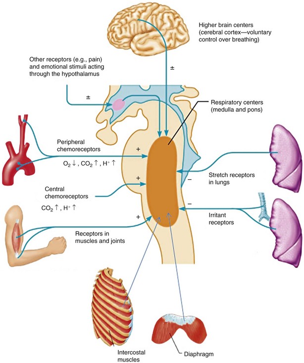
The respiratory controller receives information from a variety of sources. Some of these involve the relatively straightforward chemoreceptor signals that provide closed-loop information on the gas exchange functions of the lung. These signals arise mainly from the central and peripheral chemoreceptors that mediate the response to hypoxia, hypercapnia, and acidemia. In addition, at any given time, many other inputs from the upper airways, the lung, the respiratory muscles, and the thoracic cage may be important in determining ventilatory drive (Figure 6-2). The states of cortical arousal, sleep, and emotion play important roles in the level of ventilation and the response to other stimuli.


Peripheral Chemoreceptors


The peripheral chemoreceptors include the carotid bodies and the aortic bodies. The carotid bodies are much more important than the aortic bodies in humans. The peripheral chemoreceptors are sensitive to both hypoxia and hypercapnia or acidosis. The site of chemoreception in the carotid body is the type I glomus cells; the type II cells play more of a supporting role, similar to that of glial cells. The hypoxic response causes a sharp increase in firing rate of the carotid sinus nerve when the PaO2 is lowered below 60 mm Hg. Signal transduction involves the depolarization of the type I cells (by closing a potassium channel that normally is open at resting membrane potential). After the transduction in the type I cells, the signal is transmitted to the carotid sinus nerve endings. Rather than there being a single neurotransmitter, multiple inhibitory and excitatory neurochemicals function both as classical neurotransmitters and also as neuromodulators. Dopamine is abundant in type I cells but seems to be an inhibitory neurotransmitter. Adenosine triphosphate (ATP), by contrast, functions as the primary excitatory neurotransmitter, perhaps coreleased with acetylcholine.


Upper and Lower Respiratory Tract Receptors


Important receptors in the lung and the upper respiratory tract provide afferent information to the respiratory centers. This information is used in normal ventilation as well as to initiate maneuvers such as sneezing and coughing that need to override the gas exchanging role of the ventilatory system.


Reflexes from all along the respiratory tract provide information to the respiratory centers that will modify or sometimes even block the respiratory drive. Many of the reflexes of the airway are involved in protection, either through trying to clear the airway of foreign material through sneezing or coughing, or in preventing aspiration by closing the larynx during the swallowing of emesis. Irritant receptors are found in the nose and upper airways. They are triggered by nonspecific irritants, and their stimulation leads to reflex apnea. Pharyngeal reflexes are important in maintaining a patent airway. During inspiration, the pressure in the airway is negative, and because no intrinsic structures are present to hold the pharyngeal airway open (as with the tracheal cartilages), muscle tone must provide the counterforces to maintain an open airway. Receptors in the pharynx sense this negative pressure and signal the need for increased drive to the upper airway muscles during inspiration. In obstructive sleep apnea, this reflex may not be sufficient to overcome the forces that collapse the airway during inspiration.


Reflexes in the lower airway (tracheobronchial tree) also are involved in both shaping the ventilatory pattern and protecting the airway. Rapidly adapting pulmonary stretch receptors are so named because during constant stimulation they initially fire very rapidly but then soon decrease their firing rate. These receptors are located between airway epithelial cells and are found in abundance throughout the carina and at subsequent bronchial bifurcations. These locales are where contaminants in the inspired air (particles) are most likely to impact because of their mass. They are stimulated by irritant gases, histamine, and rapid or extreme lung inflation. They mediate reflex cough, bronchoconstriction, and hyperpnea. The slowly adapting pulmonary stretch receptors are located in airway smooth muscle and carry impulses in the vagus nerve by way of large myelinated fibers. They are activated by high lung volume or bronchoconstriction and mediate the Hering-Breuer reflex (early termination of inspiration, which in humans becomes active at an inspired volume of about 1 to 1.5 L). The J receptors, whose impulses are carried in small unmyelinated C fibers of the vagus nerve, have been so called because the nerve endings are found near (“juxta”) the alveolus in the walls of pulmonary capillaries or interstitium. They respond to mechanical deformation (e.g., pulmonary edema). Activation of these receptors causes rapid, shallow breathing and dyspnea.


The Efferent Limb: the Respiratory Muscles


Functional Anatomy


The Intercostal Muscles


The intercostal muscles are two thin layers of muscle fibers occupying each of the intercostal spaces. They are termed external and internal because of their surface relations, the external being superficial to the internal. The muscle fibers of the two layers run at approximately right angles to each other.The external intercostals extend from the tubercles of the ribs dorsally to the costochondral junctions ventrally, and their fibers are oriented obliquely, downward, and forward, from the rib above to the rib below. The internal intercostals begin posteriorly as the posterior intercostal membrane on the inner aspect of the external intercostal muscles. From approximately the angle of the rib, the internal intercostal muscles run obliquely, upward, and forward from the superior border of the rib and costal cartilage below to the floor of the subcostal groove of the rib and the edge of the costal cartilage above, ending at the sternocostal junctions. All of the intercostal muscles are innervated by the intercostal nerves.


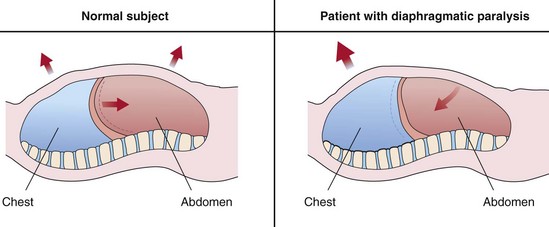
The external intercostal muscles have an inspiratory action on the rib cage, whereas the internal intercostal muscles are expiratory. An illustrative clinical example of the “isolated” inspiratory action of the intercostal muscles is offered by bilateral diaphragmatic paralysis. In patients with this deficit, inspiration is accomplished solely by the rib cage muscles. As a result, the rib cage expands during inspiration, and the pleural pressure falls. Because the diaphragm is flaccid and no transdiaphragmatic pressure can be developed, the fall in pleural pressure is transmitted to the abdomen, causing an equal fall in the abdominal pressure. Hence, the abdomen moves paradoxically inward during inspiration, opposing the inflation of the lung (Figure 6-3). This paradoxical motion is the cardinal sign of diaphragmatic paralysis on clinical examination and is invariably present in the supine posture, during which the abdominal muscles usually remain relaxed during the entire respiratory cycle. However, this sign may be absent in the erect posture.


The Diaphragm


The floor of the thoracic cavity is closed by a thin musculotendinous sheet, the diaphragm—the most important inspiratory muscle, accounting for approximately 70% of minute ventilation in normal subjects. The diaphragm is anatomically unique among the skeletal muscles in that its fibers radiate from a central tendinous structure (the central tendon) to insert peripherally into skeletal structures. The muscle of the diaphragm has two main components as defined at its point of origin: the crural (vertebral) part and the costal (sternocostal) part. The crural part arises from the crura (strong, tapering tendons attached vertically to the anterolateral aspects of the bodies and intervertebral disks of the first three lumbar vertebrae on the right and two on the left) and the three aponeurotic arcuate ligaments. The costal part of the diaphragm arises from the xiphoid process and the lower end of the sternum and the costal cartilages of the lower six ribs. These costal fibers run cranially so that they are directly apposed to the inner aspect of lower rib cage, creating a zone of apposition.


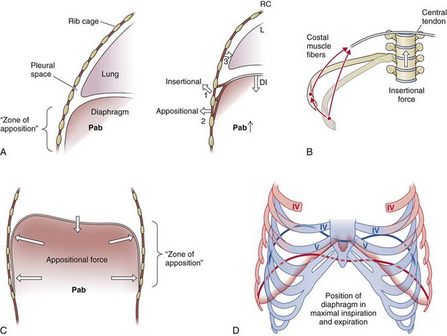
The shape of the relaxed diaphragm at the end of a normal expiration (at functional residual capacity [FRC]) is that of two domes joined by a “saddle” that runs from the sternum to the anterior surface of the spinal column (Figure 6-4). The motor innervation of the diaphragm is from the phrenic nerves, which also provide a proprioceptive supply to the muscle. When tension develops within the diaphragmatic muscle fibers, a caudally oriented force is applied on the central tendon, and the dome of the diaphragm descends; this descent has two effects. First, it expands the thoracic cavity along its craniocaudal axis, and consequently the pleural pressure falls. Second, it produces a caudal displacement of the abdominal visceral contents and an increase in the abdominal pressure, which in turn results in an outward motion of the ventral abdominal wall and the lower rib cage (appositional force). Thus, when the diaphragm contracts, a cranially oriented force is being applied by the costal diaphragmatic fibers to the upper margins of the lower six ribs that has the effect of lifting and rotating them outward (insertional force) (see Figure 6-4). The actions mediated by the changes in pleural and abdominal pressures are more complex. Viewed as the only muscle acting on the rib cage, the diaphragm has two opposing effects when it contracts: On the upper rib cage, it causes a decrease in the anteroposterior diameter, and this expiratory action results primarily from the fall in pleural pressure (see Figure 6-4). On the lower rib cage, it causes an expansion. In fact, this is the pattern of chest wall motion observed in tetraplegic patients with transection injury at the fifth cervical segment of the spinal cord or below, who have complete paralysis of the inspiratory muscles except for the diaphragm. This inspiratory action on the lower rib cage is caused by the concomitant action of two different forces, the “insertional” force already described and the “appositional” force.


Jun 12, 2016 | Posted by in RESPIRATORY | Comments Off on Control of Ventilation and Respiratory Muscles

Full access? Get Clinical Tree

Get Clinical Tree app for offline access