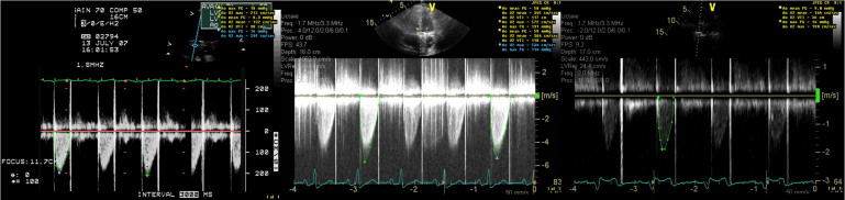
Prosthetic valve thrombosis (PVT) is a serious complication after cardiac valve replacement and usually requires urgent management with either thrombolysis or surgery. A 38-year-old woman with history of ischemic stroke and multiple valvular surgeries including aortic St. Jude mechanical valve replacement (St. Jude Medical, St. Paul, Minnesota) presented with new-onset dyspnea on exertion. Transthoracic echocardiography and 2-dimensional transesophageal echocardiography (TEE) suggested an abnormally high gradient across the aortic valve ( Figure 1 ) but could not determine the cause of the stenosis ( Figure 2 , Video 1 ). Three-dimensional (3D) TEE showed a small mobile thrombus attached to 1 disc of the St. Jude valve, resulting in partially restricted disc mobility ( Figure 2 , Videos 2 and 3 ). Also, valvular “pannus” formation was minimal, and the mobility of the other disc of the St. Jude mechanical prosthesis was normal. Thrombectomy with a carbon dioxide high-flow mister blower was chosen to eliminate the thrombus. After elimination of the thrombus and scant fibrous tissue on the underside of the valve, the mobility of the affected disc recovered completely. The gradient across valve was normalized ( Figure 1 ). The patient became asymptomatic and was discharged.
Stay updated, free articles. Join our Telegram channel

Full access? Get Clinical Tree


