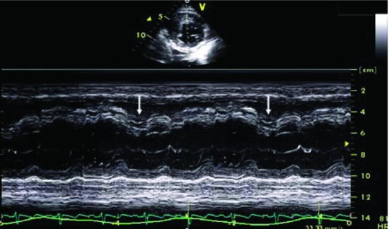
Figure 68.2 shows an M-mode recording of the septal position which varies with respiration, and during inspiration interventricular septum deviates to the left.
Figure 68.2 M-mode recording shows the septal position varies with respiration, and during inspiration, interventricular septum deviates to the left (arrows).

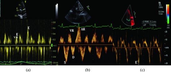
Figure 68.3 Doppler and pulse tissue Doppler recordings demonstrated: (a) a marked inspiration fall in peak mitral E-wave velocity (mean >30% from expiration). (b) A large atrial reversal wave (AR) of hepatic vein. Doppler recording indicated high-end diastolic pressure of RV. (c) Peak early longitudinal velocities (E′) in patients with constrictive pericarditis are high.
Figure 68.3 Doppler and pulse tissue Doppler recordings demonstrated: (a) a marked inspiratory fall in peak mitral E-wave velocity (mean >30% from expiration). (b) A large atrial reversal wave (AR) of hepatic vein. This Doppler recording indicated the end diastolic pressure of right ventricle is high. (c) Peak early longitudinal velocities (E′) in patients with constrictive pericarditis are high. AR, aortic regurgitation; VR, ventricular reversal.

Stay updated, free articles. Join our Telegram channel

Full access? Get Clinical Tree


