Fig. 9.1
Tubular deformity of the left ventricle in pericardial constriction
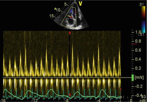
Fig. 9.2
Respiro-phasic variation due to ventricular interaction in constriction
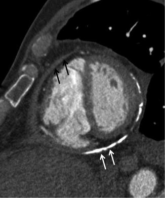
Fig. 9.3
Pericardial thickening (black arrows) and calcification (white arrows) as demonstrated by CT
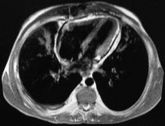
Fig. 9.4
Pericardial enhancement and circumferential thickening by MRI
Further patient information is available at:
References
1.
2.
Ling LH, Oh JK, Schaff HV, Danielson GK, Mahoney DW, Seward JB, et al. Constrictive pericarditis in the modern era: evolving clinical spectrum and impact on outcome after pericardiectomy. Circulation. 1999;100(13):1380–6. Epub 1999/09/29.PubMedCrossRef
Stay updated, free articles. Join our Telegram channel

Full access? Get Clinical Tree


