8 Congenital Heart Disease
Atrial Septal Defects
Key Points
All Atrial Septal Defects (ASD) (Tables 8-1 and 8-2)
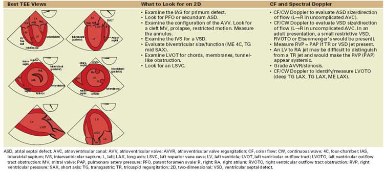
TABLE 8-1 BASIC ECHOCARDIOGRAPHIC PRINCIPLES WHEN IMAGING PATIENTS WITH AN ATRIAL SEPTAL DEFECT
asc, ascending; ASD, atrial septal defect; AV aortic valve; 4C, four-chamber; CF, color flow; IAS, interatrial septum; L, left; LAA, left atrial appendage; LSVC, left superior vena cava; LUPV, left upper pulmonary vein; ME, midesophageal; MR, mitral regurgitation; MV, mitral valve; PAP, pulmonary artery pressure; PBF, pulmonary blood flow; PS, pulmonic stenosis; PulmV, pulmonary valve; PV, pulmonary vein; PW, pulsed wave; R, right; RAE, right atrial enlargement; RV, right ventricular; RVE, right ventricular enlargement; RVH, right ventricular hypertrophy; RVP, right ventricular pressure; SAX, short axis; TR, tricuspid regurgitation; TV, tricuspid valve; 2C, two-chamber; 2D, two-dimensional; UE, upper esophageal.
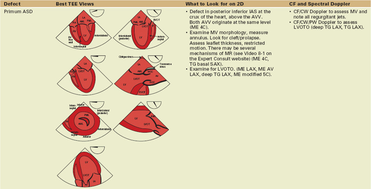
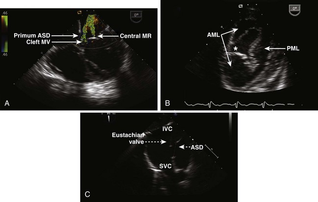
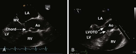
Primum Atrial Septal Defects (Figure 8-1A)
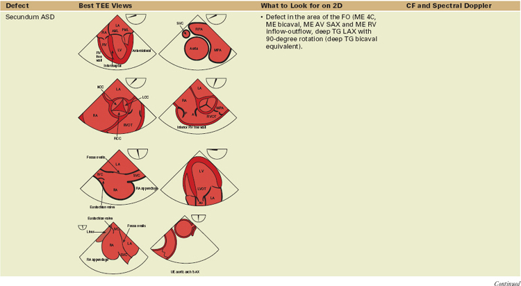
Secundum Atrial Septal Defects (see Figure 8-1C)
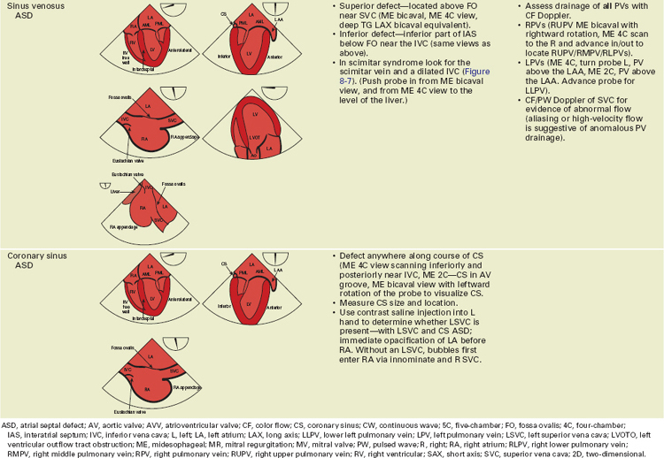
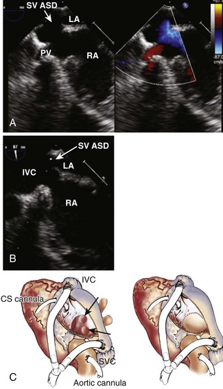
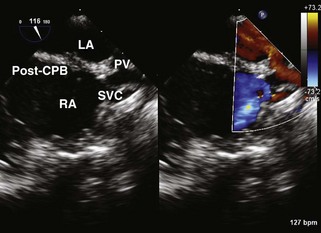
Sinus Venosus Atrial Septal Defects (Figure 8-3)
Principles of Surgical/Interventional Management
Sinus Venosus Atrial Septal Defects
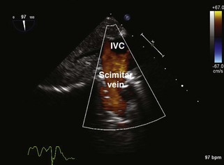
Scimitar Syndrome
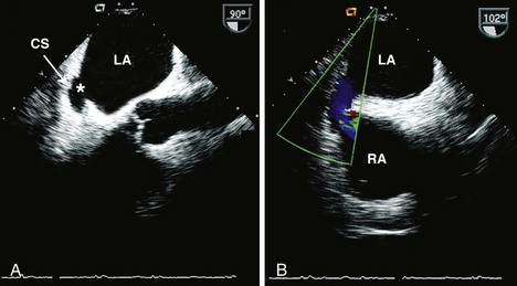
Coronary Sinus Atrial Septal Defects
Postoperative TEE Assessment after Atrial Septal Defect Repair
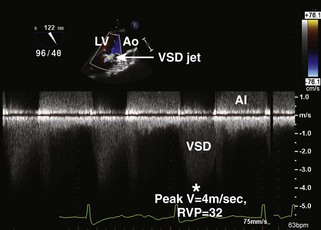
Ventricular Septal Defects
Key Points
All Ventricular Septal Defects (Tables 8-3 to 8-5)
TABLE 8-3 SURGICAL APPROACH TO VENTRICULAR SEPTAL DEFECT CLOSURE
| Most Common Surgical Approach | Defect | Additional Facts |
|---|---|---|
| RA | ||
| RV | ||
| Transpulmonary | ||
| Transaortic | ||
| LV |
DORV, double-outlet right ventricle; LV, left ventricle; LV, left ventricular; RA, right atrium; RA, right atrial; RV, right ventricle; RV, right ventricular; TOF, tetralogy of Fallot; TV, tricuspid valve; VSD, ventricular septal defect.
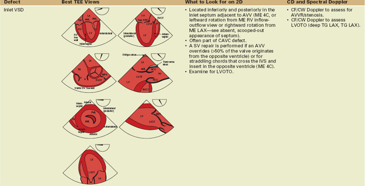
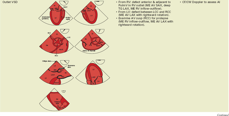
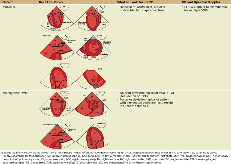
TABLE 8-4 BASIC ECHOCARDIOGRAPHIC PRINCIPLES WHEN IMAGING PATIENTS WITH A VENTRICULAR SEPTAL DEFECT
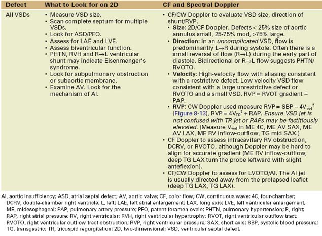
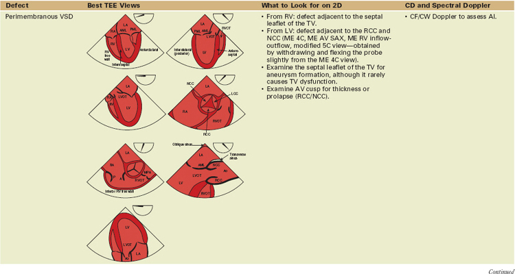
Perimembranous Defects (Figure 8-9A)
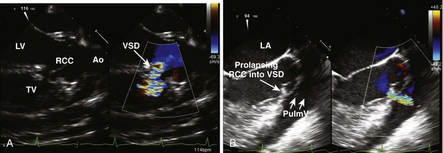
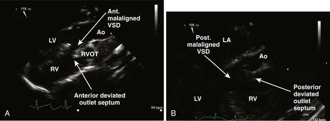
Outlet Defects (see Figure 8-9B)
Principles of Surgical Management
Postoperative TEE Assessment after Primary or Revision Ventricular Septal Defect Repair
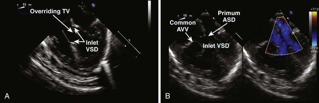
Atrioventricular Canal Defects (Table 8-6)
TABLE 8-6 BASIC ECHOCARDIOGRAPHIC PRINCIPLES WHEN IMAGING PATIENTS WITH REPAIRED OR UNREPAIRED ATRIOVENTRICULAR CANAL DEFECTS
Key Points
Transitional Canal
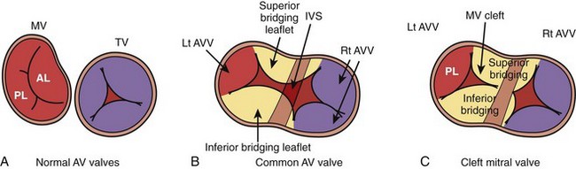
Common Atrioventricular Canal
Principles of Surgical Management
Tetralogy of Fallot
Key Points
All Tetralogy of Fallot (Tables 8-7 and 8-8)
TABLE 8-7 PRINCIPLES OF SURGICAL/INTERVENTIONAL MANAGEMENT
| Defect |
|---|


