Congenital Heart Disease


Congenital Heart Disease


Allen P. Burke, M.D.

Joseph J. Maleszewski, M.D.


General


Epidemiology

Congenital abnormalities of the heart and aortic arch (excluding the congenitally bicuspid aortic valve) occur in slightly less than 1% of the population1 and range from severe defects that result in life-threatening symptoms to incidental abnormalities. Congenital anomalies can affect multiple chambers or valves, resulting from a significant development defect (complex congenital heart disease). Isolated congenital disease of the valves or isolated atrial septal defects (ASDs) may remain asymptomatic for prolonged periods, and small ventricular septal defects may heal spontaneously. The approximate frequencies of various forms of congenital heart disease are presented in Table 140.1.


Genetics

There are several congenital syndromes associated with specific heart defects, the most common being Down syndrome and 22q11.2 deletion syndrome (see Table 140.2). In infants with heart defects, ˜20% have a defined syndrome, about half of which have gross chromosomal defects and half with defects at the gene level. Overall, up to 45% of patients with congenital heart disease have extracardiac anomalies.

Trisomy 21 (Down syndrome) is associated with heart defects in about 40% of patients and, because of its high prevalence, accounts for the largest single group of chromosome-related heart disease. The most characteristic defect is the atrioventricular (AV) septal defect (AV canal defect), but other anomalies, including ventricular septal defect, ASD, and patent ductus arteriosus (PDA), also occur with increased frequency in Down syndrome.

The 22q11.2 deletion syndrome, which includes velocardiofacial and DiGeorge syndromes, is the most frequent known chromosomal microdeletion syndrome, with an incidence of 1 in 4,000 to 5,000 live births. It is characterized by a 3-Mb deletion on chromosome 22q11.2, T-cell deficits, cleft palate facial anomalies, and hypocalcemia. Mutations in TBX1, LHX2, and TCF21 are associated with the 22q11.2 syndrome.4,6 Approximately 80% of patients have heart defects that include conotruncal anomalies, including truncus arteriosus, and anomalies of the aortic arch, including interruption of the aortic arch.4

A family history of congenital heart disease, from relatively simple to complex forms, increases the recurrence in siblings (Table 140.3). Therefore, genetics and epigenetics are likely to play a more significant role than we can directly account for at this time.


TABLE 140.1 Approximate Frequencies of Congenital Heart Diseases, as Proportion of Total


Ventricular septal defect


41%


Atrial septal defect


10%


Pulmonary valve stenosis


8%


Patent ductus arteriosus


8%


Tetralogy of Fallot


5%


Coarctation of the aorta


5%


Atrioventricular septal defect


4%


Transposition of the great arteries


4%


Aortic valve stenosisa


3%


Hypoplastic left heart


3%


Double outlet right ventricle


2%


Pulmonary atresia with VSD


1%


Pulmonary atresia with intact VSb


1%


Ebstein anomaly


1%


Double inlet left ventricle


1%


TAPVC


1%


Tricuspid atresia


1%


Other


1%


a Congenitally dysplastic stenotic valve.

b Hypoplastic right heart syndrome.


VS, ventricular septum; VSD, ventricular septal defect; TAPVC, total anomalous pulmonary venous connection.


From van der Bom T, et al. The prevalence of adult congenital heart disease, results from a systematic review and evidence based calculation. Am Heart J. 2012;164(4):568-575,


Ref. 2; Hoffman JI, Kaplan S. The incidence of congenital heart disease. J Am Coll Cardiol. 2002;39(12):1890-1900, Ref. 3.


TABLE 140.2 Syndromes and Congenital Heart Diseasea


Syndrome


Heart Defect


Percentage with Heart Involvement


Chromosomal abnormalities


Trisomy 21 (Down)


VSD, AVSD


40


Trisomy 18 (Edwards)


VSD, AVSD, PVD


95


Trisomy 13 (Patau)


DORV, ToF, VSD, ASD, PDA, PVD, Anomalous pulmonary veins


75


Monosomy X (Turner)


Coarctation, aortic stenosis


30


47,XXY (Klinefelter)


ASD, PDA, MVP


50


Partial chromosomal abnormalities (deletions and tetrasomies)


22q11.2 syndromeb (Del 22q11.2)


DiGeorge (TBX1, LHX2, TCF21): TA, ToF, PA-VSD, AAA


75-80


Velocardiofacial: ToF, TA, AAA


Williams syndrome (Del 7q11.23)


Supravalvar aortic stenosis


65


Cat eye (tetrasomy 22p)


Anomalous pulmonary veins


50


Genetic abnormalities


Noonan syndrome (PTPN11, KRAS, SOS1, and others)


Hypertrophic cardiomyopathy, PS, PVD


85


Heterotaxy (ZIC3, LEFTY2, CFC1, NODAL, ACVR2B)


Anomalous pulmonary venous connection, AVSD, VI, TGA, DORV, PS


95


Alagille (NOTCH2, JAG1)


ToF, PS, PPAS, ASD


90


Holt-Oram (TBX5)


ASD, VSD, AVSD, ToF


80


CHARGE (CHD7, SEMA3E)


ASD, PDA, MVP


65


Ellis-van Creveld (EVC, EVC2)


AVSD, ASD


65


Smith-Lemli-Opitz (DHCR7)


AVSD, partial anomalous pulmonary veins


45


aAdapted from Blue GM, et al. Congenital heart disease: current knowledge about causes and inheritance. Med J Aust. 2012;197(3):155-159, Ref. 4; Goldmuntz E, Crenshaw ML, Lin AE. Genetic aspects of congenital heart defects. In: Allen HD, et al., eds. Moss and Adams’ Heart Disease in Infants, Children, and Adolescents Including the Fetus and Young Adult. Philadelphia, PA: Lippincott Williams & Wilkins; 2013:617-643, Ref. 5. Implicated genes are in italics.

b Includes DiGeorge syndrome, velocardiofacial syndrome, Shprintzen syndrome, conotruncal anomaly face syndrome, Strong syndrome, congenital thymic aplasia. Abbreviations: ToF, tetralogy of Fallot; AAA, aortic arch anomalies; AS, aortic stenosis; ASD, atrial septal defect; AVSD, atrioventricular septal defect (primum and complete types); DORV, double outlet right ventricle; TGA, transposition of the great arteries; PA-VSD, pulmonary atresia with ventricular septal defect; PS, pulmonary stenosis; PDA, patent ductus arteriosus; PPAS, peripheral pulmonary artery stenosis; PVD, polyvalvular dysplasia; TA, truncus arteriosus; VI, ventricular inversion.


Intrauterine Risk Factors

Intrauterine rubella infection is associated with congenital heart defects in up to 45% of fetuses and neonates, including PDA, ventricular septal defect, pulmonary stenosis, and peripheral pulmonary artery stenosis.


TABLE 140.3 Recurrence Risk Siblings, Percent, After Diagnosis of Infant or Fetus with Congenital Heart Disease, by Type


• VSD


4-6


• PDA


2.5-3


• Cushion


2-3


• Coarctation


2-7


• Tetralogy


2-3


• TGA


2


• HLHS


1-4


VSD, ventricular septal defect; PDA, patent ductus arteriosus; TGA, transposition of the great arteries; HLHS, hypoplastic left heart syndrome.


Intrauterine exposure to thalidomide results in a 30% risk of congenital heart defects, including atrial and ventricular septal defects and conotruncal anomalies. Retinoic acid has similar affects, with a 25% rate of defects including ventricular septal defect and tetralogy of Fallot.


Approach to Autopsy

Congenital heart disease is generally diagnosed clinically and known at the time of autopsy. Most patients with complex congenital heart disease have undergone surgical intervention. Familiarity with the surgical procedures and clinical complications, as well as knowledge of the clinicians’ concerns, is important. Review of computed tomography angiography reports and other imaging studies and correlation of such with autopsy are critical. Often, the surgical team involved in any intervention will request attendance at the postmortem and can provide invaluable clinical information.

The pathologist may encounter untreated congenital heart disease in babies with inoperable congenital defects, often with extracardiac anomalies. Knowledge of fetal ultrasound findings is very helpful before dissection of the heart, but the pathologist should be guided by the observed anatomy and should not rely solely on imaging or intraoperative impressions.

The dissection of congenital heart specimens should accommodate answering specific questions of the clinicians, expose any abnormalities or complications, and maintain the specimen for photography and potential second opinion recognizing that few centers have the expertise to evaluate the full spectrum of congenital heart disease. Care should be taken to keep the great vessels attached. To these ends, keeping the lungs with the heart will help to document pulmonary venous connections. Thereafter, the lungs, pulmonary veins, ascending aorta, and pulmonary trunk, with the intervening ductus, can be kept as a separate block by transecting the aorta and pulmonary artery near the valves.

There are many facets to the autopsy of a child (or adult) with congenital heart disease, in addition to documenting the underlying malformation. It is important to diagnose extracardiac anomalies (which can help to establish a syndrome) and secondary changes, especially hypertensive changes in pulmonary arteries and chronic congestive changes in pulmonary and hepatic veins. Cardiac hypertrophy secondary to stenosis and shunting can be marked, and right ventricular hypertrophy is frequent in patients with pulmonary hypertension. Myocardial ischemia may occur postoperatively and should be described similar to that seen in native coronary disease. Dilatation of the ventricles and atria should be described.

Interventions (aided by review of the operative notes and imaging studies), including anastomoses, prior takedowns (especially systemic-pulmonary arterial shunts), anastomotic strictures, thrombosis, or narrowing of shunts, are all to be evaluated and documented. It is helpful to review the operative notes prior to autopsy and familiarize oneself with the specific procedure performed.


General Approach and Segmental Analysis

Specimens to be evaluated for congenital heart disease should be approached in a uniform and consistent fashion. Such evaluation begins with a description of the location and position of the heart within the chest, then a thorough description of all the connections (venoatrial, atrioventricular, and ventriculoarterial), and then documentation of any abnormal shunts.


Cardiac Position and Sidedness (Situs)

The normal location (or position) of the heart in the left hemithorax is termed “levoposition.” Dextroposition (displacement into the right hemithorax) or mesoposition (midline displacement) often result from extracardiac causes, for example, pulmonary hypoplasia or scoliosis. The direction of the apex is usually described separately as leftward (normal), rightward, or midline.7

There are three asymmetric organs: the heart, lungs, and gut. The spleen is a unilateral organ that is left sided from inception. The situs of the three organs indicates the status of normally manifest sidedness: normal (situs solitus), mirror image (situs inversus), right isomerism
(both sides have typical right-sided features), and left isomerism (both sides have typical left-sided features). The spleen, being left sided, is absent in right-sided isomerism, on the right in situs inversus, and multiple in left-sided isomerism.
The sidedness (situs) of the heart is determined solely by atrial morphology and can be described as normal (solitus), inverted (inversus), or duplicated (ambiguous). Right atrial features include the presence of the following: crista terminalis, coronary sinus ostium, limbus of the oval fossa, pyramidal atrial appendage, and a suprahepatic portion of the inferior vena cava. Left atrial morphology is defined by a lack of conspicuous pectinate muscles and a narrow-based atrial appendage with variable morphology.

Like atrial morphology, ventricular morphology is side specific. As a rule, the atrioventricular valves always ride with their corresponding ventricle. The inflow, apical trabeculated, and outflow portions of the ventricle each have unique features. The inflow of the morphologic right ventricle (RV) has an atrioventricular (tricuspid) valve with direct septal chordal insertions. The apical trabeculations of a morphologic RV are more coarse than those observed in a morphologic left ventricle (LV), and the outflow portion of the right ventricle is muscular. All tendinous cords on the left are attached to papillary muscles (without direct septal chordal insertions). The apex has finer trabeculations than those seen in a morphologic RV, and the outflow tract is musculomembranous owing to the continuity between the atrioventricular and semilunar valves.

The sidedness of the heart, lungs, and abdominal viscera is not always the same. In complete situs inversus, all three organs are inverted. However, in some cases of heterotaxy syndromes, the heart and possibly lungs may be the only organs involved. Pulmonary sidedness is defined by the relationship of the pulmonary arteries to the upper lobe bronchus. The right side pulmonary artery is normally anterior to the upper lobe bronchus and the left side superior to the upper lobe bronchus. Number of lobes (three on the right, two on the left) is not a reliable indicator of sidedness.


Venoatrial Connections

The inferior vena cava normally enters the right atrium. It may enter the left side of a common atrium or left-sided atrium in heterotaxy syndromes. It may also end in the liver, with azygous connection to the atria. The interrupted inferior vena cava syndrome is associated with heart defects in 85% of cases, especially polysplenia.8

The normal left-sided venoatrial connections consist of pulmonary veins (usually two left and two right) emptying into the chamber. Variability in number is not uncommon, but all connections should be documented.


Atrioventricular Connections

Atrioventricular connections can be described as concordant (morphologic atrium to respective morphologic ventricle) or discordant. Ventricular inversion (atrioventricular discordance) may be encountered and is the hallmark of congenitally corrected transposition of the great arteries (L-TGA). Univentricular connections may be the result of a single connection (e.g., tricuspid atresia, where there is absent right-sided and normal left-sided connection); double inlet ventricle (usually left), where two mirror-image mitral-like valves enter the ventricle, with a rudimentary right ventricle; or common inlet atrioventricular valve into a single ventricle (usually right), with a rudimentary left ventricle. The latter condition is highly associated with asplenia.


Ventriculoarterial Connections

Ventriculoarterial discordance is more common than atrioventricular discordance and is seen in all cases of transposition of the great arteries (both D and L types). In this condition, the morphologic right ventricle gives rise to the aorta and the morphologic left ventricle the pulmonary artery. The semilunar valve always accompanies the artery; therefore, the aortic valve is present in the artery that gives rise to the coronary and arch vessels and the pulmonary valve the artery that supplies the lungs.7

Other abnormal connections include double outlet ventricles (rare and more common on the right), single outlet (pulmonary atresia or hypoplastic left heart syndrome), and common outlet ventricles (truncus arteriosus).


Valvular Atresia, Stenosis, and Absence

The term “atresia” indicates lack of connection across two chambers normally connected through a valvular orifice. Stenosis indicates decreased flow with a gradient. Absent valve indicates no valve leaflets, but a widely patent outflow (seen exclusively in the pulmonary position in variants of tetralogy of Fallot, where there is marked dilatation of the annulus).


Shunts

A shunt is defined as an abnormal connection between two noncontiguous cardiac structures resulting in abnormal blood flow. The most common type of shunt is a left-to-right shunt (e.g., ASD, ventricular septal defect, patent ductal artery), which results in blood traveling from the systemic circulation into the pulmonary circulation. Clinically, untreated septal defects result in progressive heart failure and acyanotic heart disease, until there is reversal of flow and cyanosis ensues.


Aortic Arch Anomalies


Embryology and Interruption of the Aortic Arch

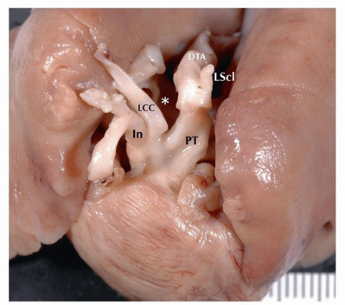
The dorsal aortas are initially connected to the aortic sac by three sets of arteries that originate from the pharyngeal arches III, IV, and VI. Those from arch III become the carotid and subclavian arteries, IV the aortic arch and innominate artery, and VI the pulmonary arteries and ductus. The ascending aorta and the C segment of the aorta (from the innominate to the left common carotid) are derived from the aortic sac, the B segment from the IV arch (left common carotid to left subclavian), and the A segment (isthmus) from the dorsal aorta. The numbers A, B, and C refer not only to the segments of the aorta but also to the sites of interruption in congenitally interrupted aortic arch. Type B, for example, is the most common seen in DiGeorge syndrome (Fig. 140.1).


FIGURE 140.1 ▲ Interrupted aortic arch type B. LCC, left common carotid; PT, pulmonary trunk; LScl, left subclavian; DTA, descending thoracic aorta. Asterisk is the location of normal arch, which is absent. Interruption between left subclavian and left common carotid is in the B aortic segment, between the left common carotid and left subclavian.


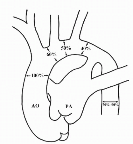
FIGURE 140.2 ▲ Normal diameters of the aortic arch in utero. Note that the isthmus is normally 40% that of the ascending aorta; narrowing of the isthmus should not be overcalled as coarctation. Reproduce with permission, Siewers RD, Ettedgui J, Pahl E, et al. Coarctation and hypoplasia of the aortic arch: will the arch grow? Ann Thorac Surg. 1991;52:608-614.


Coarctation of the Aorta

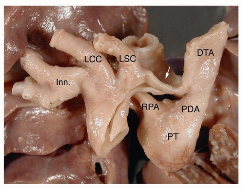
Aortic coarctation usually occurs at the level of the left subclavian artery (distal arch) (Figs. 140.2, 140.3, 140.4). In severe infantile coarctation, aortic flow is dependent on ductal flow (“preductal coarctation”). Less severe forms present as hypertension in children and adolescents and are often “postductal.”

Most cases of congenital coarctation of the aorta are associated with other cardiac defects, usually congenital left-sided obstruction such as the bicuspid aortic valve, which is seen in approximately half of cases of aortic coarctation. The association of coarctation with supravalvar mitral ring, parachute mitral valve, and subaortic stenosis is termed “Shone complex.”9 There is also an association with cerebral aneurysms and Marfan syndrome.


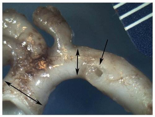
FIGURE 140.3 ▲ Normal infant aortic arch. Double arrows show ascending aorta (left) and isthmus (right). Single arrow shows the orifice of the ductus arteriosus.


FIGURE 140.4 ▲ Aortic coarctation, preductal. The arrow points to the coarctation at the isthmus. Inn, innominate; LCC, left common carotid; LSC, left subclavian; RPA, right pulmonary artery; PT, pulmonary trunk; PDA, patent ductus arteriosus; DTA, descending thoracic aorta.

Surgical corrections of coarctation include balloon angioplasty and resection with placement of an interposition graft. Complications of balloon angioplasty include restenosis, postdilatation aneurysms for native coarctation, aortic dissection, and aortic perforation. Surgical repair of coarctation includes the subclavian flap, which sacrifices the subclavian artery. For this reason, it is recommended only for patients under 1 year of age, so that collateral may form. End-to-end anastomotic repair may be performed in older patients. Complications include recurrent stenosis, persistent left ventricular hypertrophy, and aneurysms at site of repair, especially with patch aortoplasty. Patch repair may be complicated by rupture and endocarditis.


Double Aortic Arch

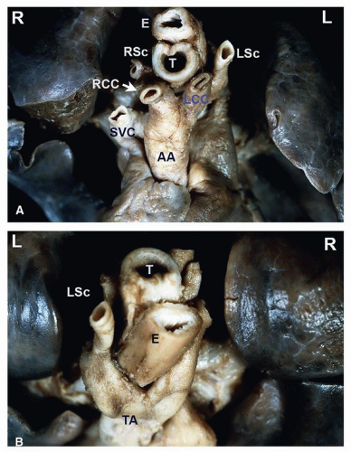
In double aortic arch, there is duplication of the arch around the trachea and esophagus, forming a ring. The right arch is usually larger, with a left-sided ductus arteriosus (Fig. 140.5). It is commonly isolated but can be associated with transposition of the great arteries, ventricular septal defect, persistent truncus arteriosus, and tetralogy. Double arches are typically symptomatic, with findings of stridor, dysphagia, cough, and pneumonia. Imaging studies demonstrate esophageal and tracheal compression. Diagnosis is confirmed by magnetic resonance angiography.


Aberrant (Retroesophageal) Right Subclavian Artery

Aberrant right subclavian artery is a benign isolated condition affecting 0.5% of the population. The artery arises distal to left subclavian and travels posterior to the esophagus. Oftentimes, a saccular outpouching (diverticulum of Kommerell) may be seen at the takeoff of the right subclavian from the aorta. It rarely causes symptoms, usually in the form of dysphagia (“dysphagia lusoria”). When associated with coarctation, it can cause the subclavian steal syndrome.


Right Aortic Arch

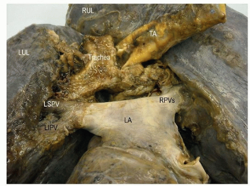
Laterality of the aortic arch is determined by the bronchus over which it travels (left bronchus, left aortic arch; right bronchus, right aortic arch) (Fig. 140.6). Typically, the branching pattern of vessels will be mirror image, such that the innominate arises first giving rise to the left subclavian and common carotid, followed by the right common carotid and right subclavian in turn. Between 20% and 30% of patients with
tetralogy, persistent truncus arteriosus, and pulmonary atresia and ventricular septal defect have right-sided arches with mirror-image branching (see below), in addition to lower rates for double outlet right ventricle, tricuspid atresia, and transposition.


FIGURE 140.5 ▲ Double aortic arch, anterior/cranial view. A. The ascending aorta bifurcates into an anterior left branch, supplying the left common carotid artery and the left subclavian artery, and a posterior right branch, supplying the right common carotid and right subclavian arteries. B. Double aortic arch, posterior/cranial view. The continuation of the aorta viewed from behind demonstrates the anterior left branch wrapping around the trachea and esophagus as well as the right posterior branch emerging from under the esophagus. The distal aorta continues as a centrally located structure. Abbreviations: RCC, right common carotid artery; LCC, left common carotid artery; RSc, right subclavian artery; LSc, left subclavian artery; E, esophagus; T, trachea; AA, ascending aorta; TA, descending aorta; SVC, superior vena cava.


FIGURE 140.6 ▲ Right aortic arch, posterior view. The descending thoracic aorta (TA) is to the right of the trachea. RUL, right upper lobe; LUL, left upper lobe; LSPV, left superior pulmonary vein; RPV, right pulmonary vein; LA, left atrium.


Abnormal Shunts


Patent Foramen Ovale

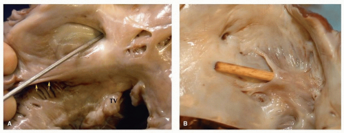
Patent foramen ovale (PFO) refers to failed fusion between the valve and limbus of the fossa ovalis, resulting in a potential interatrial communication (Figs. 140.7 and 140.8). It is evaluated for during life via echocardiography (with a bubble study) or at autopsy by the ability to pass a probe from the right atria to the left across the valve of the fossa ovalis. Its incidence is indirectly related with age, seen in approximately one in three individuals <30 years, one in four individuals between the ages of 30 and 80, and one in five individuals >80 years.

Most cases of PFO do not come to clinical attention and are incidental findings on imaging or autopsy. A small proportion, however, present with cerebrovascular symptoms of paradoxical small emboli
that may range from sporadic migraine to transient ischemic attacks or stroke. Transthoracic echocardiography with contrast or transesophageal echocardiography is usually required to detect small PFO.
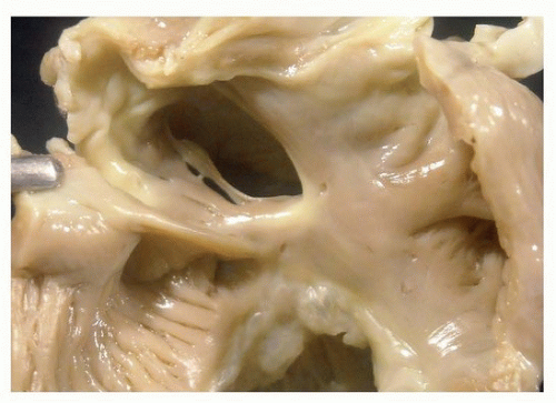


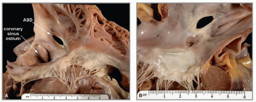
FIGURE 140.7A. Atrial septal defect, secundum type, from right atrium. The atrial septal defect (ASD) is in the area of the oval fossa. The coronary sinus ostium appears dilated. B. Atrial septal defect, secundum type, from left atrium. This defect is the same as that illustrated in figure 8, from the left side. The anterior leaflet of the mitral valve is below.


FIGURE 140.8 ▲ Patent foramen ovale, adult. A. The probe is present within the right side of the oval fossa. The eustachian valve is present at the bottom above the orifice of the coronary sinus. The septal leaflet of the tricuspid valve (TV) is present below. B. Viewed from the left, this patency was a tunnel within the foramen ovale. The characteristic “backward C” is present where the probe emerges.

In cases of atrial dilatation, stretching of the atrial wall may cause the limbus and valve portions of a PFO to pull away from one another, resulting in what looks like an ASD (so-called acquired atrial septal defect). Therefore, in cases of atrial dilatation, care should be taken to not overcall the presence of an ASD.


Patent Ductus Arteriosus

Normally, the ductal artery functionally closes within 24 hours of birth and anatomically seals within the first few months of life. PDA refers to persistent communication between the pulmonary artery and aorta through the ductal artery, allowing for a left-to-right shunt early on, which may eventually reverse and become right to left.

In utero and at birth, the duct diameter is approximate to that of the pulmonary trunk and descending thoracic aorta; if significantly narrowed in stillborns, the diagnosis of premature closure of the duct should be considered. In adulthood, calcification and aneurysms may form of the patent duct as well as endocarditis (rarely).10

If isolated, it is usually repaired without sequelae. Surgical corrections of the ductus arteriosus include open ligation, vascular clipping, transection with oversewing, and pericardial patching if there are residual defects in the aorta of pulmonary artery or ductal calcification in older patients. Percutaneous closure with device is the current procedure of choice in many centers.11


Atrial Septal Defects

ASD refers to an abnormal communication between the atria owing to a deficiency in the atrial septum. They can be categorized into one of four varieties: secundum, coronary sinus, sinus venosus, and primum. The latter, primum type, is usually considered an atrioventricular septal defect and is therefore discussed under that heading.

The most common ASD is the secundum type, representing 70% of cases. The defect is present in the region of the fossa ovalis and is thought to be secondary to incomplete formation of septum secundum or too large of an ostium secundum (Figs. 140.9 and 140.10). The flap valve may be completely absent, or partly absent with fenestrations.

Sinus venosus ASDs are rare and usually associated with partial anomalous pulmonary venous return, because of the location near the superior vena cava and the right pulmonary veins (Fig. 140.11). The location is posterior to the fossa ovalis, on the superior surface of the septum. Sinus venosus defects are often seen in association with anomalous drainage of right pulmonary veins into the right atrium (partial anomalous venous return).

The least common type of ASD is the coronary sinus type (so-called unroofed coronary sinus). Absence of the anterior portion of the coronary sinus as it enters the right atrium can result in connection to the adjacent left atrium and led to shunting of blood from the left atrium into the coronary sinus and subsequently into the right atrium. This type is often associated with a persistent left-sided superior vena cava.12

ASDs are more common in women than men with a 2 to 3:1 ratio. They are occasionally inherited.13 Symptoms of ASDs result from leftto-right shunting with resultant increased pulmonary blood flow relative to systemic flow. Patients may present with dyspnea, fatigue, and palpitations. Sizeable defects may develop into severe hemodynamic consequences that warrant surgical treatment.

Uncommonly, ASDs may be specific causes of death that are related to atrial fibrillation, right heart failure, and pulmonary hypertension. Echocardiography reliably identifies most cases of ASDs. Cardiac catheterization is used in a few cases, when assessment of hemodynamics is required.14 Surgery early in life dramatically increases life survival, especially if undertaken before the third decade of life.15 Open surgical treatment is gradually being replaced by percutaneous closure by clamshell and other types of devices.16


FIGURE 140.9 ▲ Atrial septal defect, secundum type, from the right. There is no septal tissue present in the oval fossa area, save a thin cord. The coronary sinus is present just below (posteriorly).


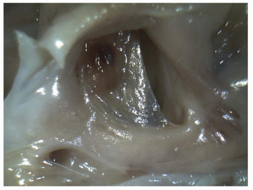
FIGURE 140.10 ▲ Patent foramen ovale, viewed from the right atrium. In this premature neonate, this is a normal finding. The oval fossa nearly covered by a thin, translucent membrane. It is easy to disrupt the ostium primum in fetal and neonatal hearts, and one should be careful not to overcall atrial septal defect.


Atrioventricular Septal Defect

Atrioventricular septal defects (AVSDs) (also called atrioventricular cushion defects) result from a defect in the region of the atrioventricular septum. By virtue of the deficient AV septum, the mitral valve is apically displaced to reside in the same plane as that of the tricuspid valve. AVSDs can be further categorized into complete and partial forms.

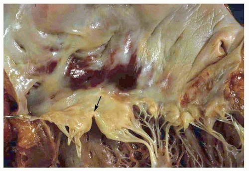
A partial AVSD indicates atrial septal involvement (so-called primum-type ASD) with separate mitral and tricuspid valve orifices. The mitral valve usually exhibits a cleft anterior leaflet (Fig. 140.12). There is no direct interventricular or atrioventricular communication. A complete AVSD results in a common atrioventricular valve, with five distinct leaflets, and direct interventricular and atrioventricular communication (Figs. 140.13, 140.14, 140.15, 140.16).


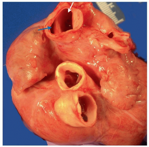
FIGURE 140.11 ▲ Sinus venosus atrial septal defect. The superior vena cava (above) is transected, revealing the superior rim of the atrial septal defect, exposing the right atrium (blue arrow) and left atrium (white arrow).


FIGURE 140.12 ▲ Partial atrioventricular canal defect. Note cleft of the anterior mitral valve leaflet (arrow).

Associated anomalies include Down syndrome (75%), tetralogy of Fallot (almost 100% association with Down syndrome), single papillary muscle in the left ventricle (important to identify prior to surgery), double outlet right ventricle, transposition of great arteries, and hypoplastic left ventricle (unbalanced AVSD).

Surgical correction involves one or two synthetic patches to cover the defect, with suspension of mitral component to the patch and valve reconstruction/replacement. The cleft in the mitral valve (present in partial AV canal) is repaired if large. Postoperative complications include mitral valve stenosis (especially if single papillary muscle or double orifice mitral valve), mitral insufficiency, residual shunts, and arrhythmias in 25% of patients, a minority of whom require pacemakers. Progressive pulmonary hypertension is especially common if there is high pulmonary resistance preoperatively, which occurs most commonly in Down syndrome.


Ventricular Septal Defect

There are several classifications for ventricular septal defects (VSDs). The simplest includes perimembranous (adjacent to the membranous septum), muscular (generally trabeculated portion of the inlet ventricle), outflow or subpulmonic, and inlet/septal (atrioventricular canal defects).12

Perimembranous ventricular septal defects account for about 35% of clinically detected VSDs (Figs. 140.17 and 140.18). Synonyms include paramembranous, infracristal, and conoventricular defects. The posterior border is the membranous septum, with the aortic and tricuspid valves in direct continuity. In some hearts, there is a muscular band that separates the defect from membranous septum. The perimembranous defects have been subclassified by the direction to which they extend, including inlet, trabecular, and infundibular.

Only gold members can continue reading. Log In or Register to continue

Stay updated, free articles. Join our Telegram channel

Aug 19, 2016 | Posted by in CARDIOLOGY | Comments Off on Congenital Heart Disease

Full access? Get Clinical Tree
Get Clinical Tree app for offline access