Figure 32.2 Apical 4-chamber view clarifies the location of the large papillary muscle and its proximity to the left ventricle septum. Note that the mitral valve stenosis obstruction (arrow) is located more towards the mid-left ventricle as compared to rheumatic mitral valve stenosis (a). Systolic color flow convergence (arrow) develops at the mid-left ventricle below the left ventricle outflow obstruction. Mitral regurgitation is evident but a significant portion of the regurgitant jet is shadowed by the large papillary muscle (b).
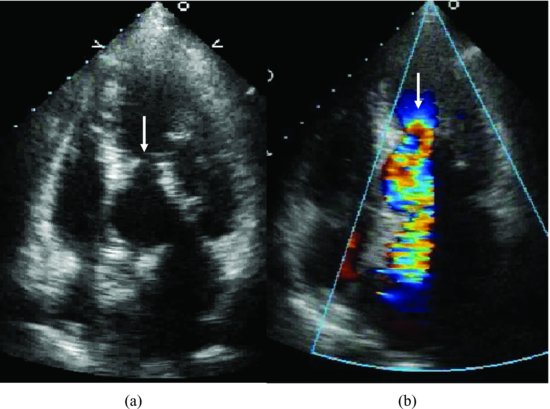
Figure 32.3 Continuous wave Doppler of mitral flow demonstrates both mitral regurgitation with a left ventricle–left atrium gradient of 140 mmHg and mitral valve stenosis with a mean gradient of 14 mmHg.
Stay updated, free articles. Join our Telegram channel

Full access? Get Clinical Tree


