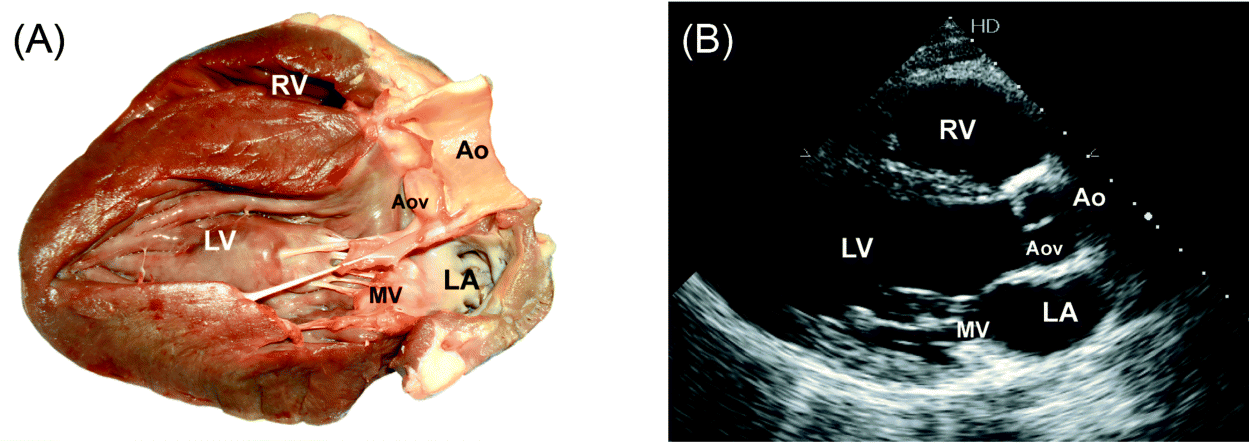
Parasternal Long-axis view (Fig. 1)
1. 2D image (4 beats) (measure LVOT diameter).
2. Color Doppler through MV and AoV (4 beats).
3. M-mode through Aortic root (measure root and LA diameter, and aortic cusp separation).
4. Color Doppler M-mode through the aortic root (4 beats).
5. M-mode through MV (± Valsalva test to check for MV prolapse and SAM; measure “E” to “S” separation distance).
6. Color Doppler M-mode through MV (4 beats).
7. M-mode through mid LV (4 beats) (measure septal and inferolateral wall thickness LVEDD and LVESD).
RA/RV view
From parasternal LAX view tilt transducer to point it to right hip:
1. 2D image (4 beats);
2. Color Doppler through TV (4 beats);
3. CW Doppler through TV to measure max TR velocity if TR jet present.
Parasternal Short-axis view (Fig. 2 and Fig. 3)
1. 2D through AoV (4 beats) (to assess structure and mobility; use zoom).
2. Color Doppler through AoV (4 beats).
3. 2D through PV (4beats).
4. Color Doppler through PV (4 beats).
5. PW Doppler at the tips of PV to measure PAT (4 beats).
6. CW Doppler through PV to measure PR velocity if present, and maximum outflow velocity through PV.
7. 2D through TV (4 beats).
8. Color Doppler through TV (4 beats).
9. PW Doppler at the tips of TV leaflets to assess inflow pattern (4 beats).
10. If inflow jet max velocity is >1.5 m/sec, trace the diastolic flow to measure mean transvalvular gradient.
11. CW Doppler through TV to measure max TR velocity if TR jet present.


