Coagulation Testing


Coagulation Testing


Jay C. Horrow

Josef Nile Mueksch

Nicholas Weber

Michael S. Green


No coagulation test can duplicate the complex milieu present at an injured vessel. Merely placing a needle or a catheter in a vessel initiates a multitude of hemostatic responses that alter measurements on the blood sample that has been removed. Surface activation of the clotting cascade begins when blood leaves the protective environment of the endothelial cells and enters into collection tubes. For these reasons, coagulation tests must be viewed as an approximation of actual events. This chapter examines the role of coagulation tests performed at central laboratory facilities and the changing landscape brought about by point-of-care testing.


CENTRALIZED COAGULATION LABORATORY TESTS

Devices using formation of a clot as an endpoint usually employ electrical or optical detection methods. The classic instrument, the Fibrometer (BBL Microbiological Systems, Cockeysville, MD), places a stationary and a moving probe in the sample. When fibrin strands bridge the two electrodes, conductivity increases causing a timer to halt (1). Laboratory prothrombin time (PT), activated partial thromboplastin time (aPTT), fibrinogen, and thrombin time determinations use this technology.


Prothrombin Time and Activated Partial Thromboplastin Time

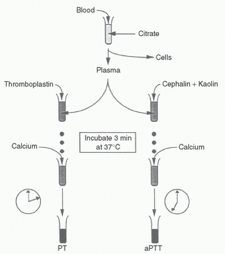
Figure 18.1 illustrates the steps in performing these tests. Each test involves centrifuging a citrated specimen of blood. Citrate complexes calcium, a necessary cofactor in the coagulation cascade, thereby preventing coagulation factor activation. The supernatant obtained by centrifugation, plasma, is incubated for 3 minutes with an additive that differs for the two tests. After addition of excess calcium with respect to citrate, the time to formation of gel is measured.

The PT, better called a complete thromboplastin time, incubates plasma with tissue extract (thromboplastin). As a tissue product, thromboplastin supplies its own phospholipid plus tissue factor. If the plasma sample contains sufficient factor VII and factors in the common pathway, it will gel in approximately 12 seconds. Variations in the potency and quality of thromboplastin reagents require simultaneous determination of a control sample and comparison of the patient’s result with the control result. Each batch of thromboplastin reagent is graded in potency. The international normalized ratio (INR) calculation allows for comparison of PT results from different commercial thromboplastin reagents: INR = PCRISI, where PCR is the ratio of patient sample to control sample PT results and ISI is the international sensitivity index, a measure of responsiveness to decreased concentrations of vitamin K-dependent factors. The higher the ISI of a thromboplastin, the less responsive the clotting time to changes in coagulation factors when using that thromboplastin. Therefore, a less responsive thromboplastin, which yields a smaller PCR, carries a larger ISI, yielding a similar calculated INR.
Thromboplastin reagent manufacturers determine the ISI by comparing the reactivity of each specific lot of thromboplastin with an international reference preparation (2).


FIGURE 18.1. Steps in performing the prothrombin time (PT, left side) and the activated partial thromboplastin time (aPTT, right side). See text for details.

The partial thromboplastin time (PTT) incubates plasma with an extract of thromboplastin that contains the phospholipid but not the tissue factor, thereby preventing activation of extrinsic factor VII. Now entirely dependent on surface activation alone, the gel forms slowly (73 ± 11 [SD] seconds) (3). The aPTT uses a surface activation accelerator such as kaolin, ellagic acid, silica, bentonite, or celite, which allows gel formation to then occur in approximately 32 seconds (1). As with the PT, simultaneous controls are required, with abnormal results at 1.5 or more times the control.

Warfarin therapy, which inhibits carboxylation of the vitamin K-dependent clotting factors, prolongs the PT because factor VII production is most vulnerable to lack of vitamin K. Heparin primarily affects the aPTT but not the PT because the potent procoagulant action of the thromboplastin reagent in the PT test easily overwhelms any inhibition of factors Xa and thrombin from the moderate doses of heparin used to treat venous thrombosis or acute coronary syndrome. The less potent partial thromboplastin reagent in the aPTT test, however, is sensitive enough to demonstrate heparin’s anticoagulant effects. Large doses of heparin will prolong the PT also.


Thrombin Time

Adding thrombin to a plasma sample will form fibrin within 10 seconds if functionally active fibrinogen is present, heparin is absent, and fibrin degradation products (FDPs) are absent. Sensitivity of the thrombin time to small amounts of heparin occurs because small amounts of thrombin are added. The traditional thrombin time is relatively insensitive to fibrinogen deficiency and to FDPs: detectable prolongation requires less than 0.75 g/L fibrinogen (4) or more than 200 mg/mL FDPs. Sensitivity to FDPs increases if fibrinogen concentrations are low. Also, diluted plasma increases sensitivity of the thrombin test to fibrinogen deficiency. Amyloidosis prolongs the thrombin time by inhibiting conversion of fibrinogen to fibrin (5).


Reptilase Time

The Reptilase time measures the interval between addition to plasma of venom from the South American pit viper Bothrops jararaca and formation of fibrin: it is like a thrombin time, but uses the venom instead of thrombin to form fibrin. A prolonged Reptilase time implicates reduced or dysfunctional fibrinogen or high concentrations of FDPs as the cause of a prolonged thrombin time. Neither heparin nor direct thrombin inhibitors prolong the Reptilase time (6). It is several-fold more insensitive to FDPs than the thrombin time. Amyloidosis also prolongs the Reptilase time.


Fibrinogen

Most laboratories use the Clauss method for fibrinogen determination, in which a thrombin time is performed on diluted plasma. With diluted samples, fibrinogen becomes the factor limiting clot formation, so that the clotting time varies inversely with fibrinogen activity (1). The Ellis method employs undiluted plasma, smaller amounts of thrombin, and a spectrophotometric measure of turbidity. A PT-based method adds thromboplastin to undiluted plasma, thereby using endogenously generated thrombin. Antibody-based tests for fibrinogen can distinguish among the dysfibrinogenemias. Decreased fibrinogen concentration occurs in end-stage hepatic failure, consumptive coagulopathy, and uncontrolled fibrinolysis leading to fibrinogenolysis, extreme hemodilution, and massive transfusion.


Fibrin Degradation Products

Immunologic tests provide a semiquantitative determination of the various fragments resulting from fibrin and fibrinogen degradation. The most common test for FDPs uses latex agglutination of serum. Note that serum, not plasma, is used. It provides results as either negative (<10 µg/mL) or positive (>10 µg/mL) for antigens, which are the breakdown products of either fibrin or fibrinogen. Serial dilutions of plasma yield more specific information when the undiluted sample is positive. More expensive quantitative analysis reveals normal serum levels of 2.1 to 2.7 µg/mL for FDPs in the absence of exercise or stress (1). This test does not differentiate the breakdown products of fibrinogen from those of fibrin.


D-Dimer

Molecules of two linked “D” domains, shown in Figure 18.2, a specific degradation product of cross-linked fibrin, can be detected either semiquantitatively with a latex agglutination technique or in a fully quantitative manner with an enzyme-linked immunosorbent assay (ELISA). Many hospital coagulation laboratories offer this test on a batched basis. The D-dimer is more specific for secondary fibrinolysis than the FDP test. Like the FDP test, D-dimer results appear as fibrinogen equivalent units (i.e., the quantity of fibrinogen initially present that leads to the observed level of breakdown product). Normally, D-dimer is less than 0.5 µg/mL (fibrinogen equivalent units). Antibody-based tests use plasma, rather than serum, because the specificity afforded by the antibody method prevents fibrinogen in plasma from confounding the results.


Platelet Count

The central role of platelets in coagulation and the impact of bypass on platelet function augment the importance of monitoring platelets during surgery. Because bypass affects both platelet function and count, measurement of platelet count is necessary but not sufficient to assess platelet role in coagulation. Although cell counters can and have been made mobile (7), measurement of platelet count has remained a central laboratory function at nearly all centers.


FIGURE 18.2. Formation of fibrin degradation products from cross-linked fibrin. Plasmin cleaves fibrin between its D and E domains at the dashed lines to yield D-dimer (DD), fragment Y (DE), fragment X (DED), and larger combinations (DY, YY, DXD, and others not shown). D-Dimer serves as a specific marker for lysis of cross-linked fibrin. (From Francis CW, Marder VJ. Physiologic regulation and pathologic disorders of fibrinolysis. In: Colman RW, Hirsh J, Marder VJ, et al., eds. Hemostasis and thrombosis. 3rd ed. Philadelphia, PA: JB Lippincott Co, 1994:1076-1103, with permission.)


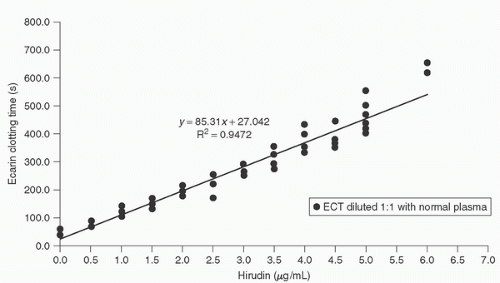
FIGURE 18.3. The dose-response curve of ecarin clotting time (ECT) to in vitro titration in six patients before cardiac surgery, with each solid dot representing results from one patient. (From Nuttall GA, Oliver WC Jr. Patients with a history of type II heparin-induced thrombocytopenia with thrombosis requiring cardiac surgery with cardiopulmonary bypass: a prospective observational case series. Anesth Analg 2003;96(2):344-350.)


Platelet Aggregometry

Platelet aggregometry utilizes a photo-optical instrument to measure light transmittance through a platelet-rich plasma sample (8). Upon exposure to a platelet agonist, the initially turbid sample shows increased light transmittance as platelets adhere to surfaces and one another. Impaired aggregation correlates poorly with clinical bleeding (8,9). Agonist agents include collagen, epinephrine, and adenosine diphosphate (ADP). Owing to the technical expertise often required to perform aggregometry, it finds application in research more often than in routine clinical care.


Ecarin Clotting Time

The injectable direct thrombin inhibitors hirudin, bivalirudin (Angiomax), and argatroban are alternatives to heparin available to patients with heparin-induced thrombocytopenia (HIT) (see Chapter 19). The ecarin clotting time (ECT) monitors the extent of anticoagulation from direct thrombin inhibitors. It is based on the conversion of thrombin to meizothrombin by ecarin, venom from the snake Echis carinatus (10). Less procoagulant than thrombin, meizothrombin binds to direct thrombin inhibitors in equimolar concentrations creating a linear correlation between ECT prolongation and thrombin inhibitor concentration (Fig. 18.3). With a point-of-care ECT test (11,12,13,14) no longer available, each hospital laboratory must create its own standardized version, thereby making the ECT extremely difficult to apply to the operative care of patients with HIT. Clinicians report successful cardiopulmonary bypass (CPB) using individual calibration curves for each patient (15). An alternative anticoagulation management scheme for patients with HIT takes the functional approach using either the activated clotting time (ACT) or aPTT,
as opposed to targeting specific blood concentrations using the ECT. Only case reports exist regarding monitoring failure or success (16,17,18,19,20,21).


POINT-OF-CARE COAGULATION TESTS

For clinicians, the ability to obtain coagulation results in a timely manner critically impacts the diagnosis and treatment of specific hemostatic derangements: results must be timely as well as accurate. Prompt turnaround potentially saves time, which translates into financial savings. The desire for accurate and prompt coagulation information has created an interest in point-of-care testing (22).

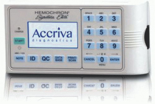
Point-of-care, also designated as “near-patient,” “on-site,” “alternate site,” or “bedside,” tests utilize whole-blood samples in close proximity to the patient, compared with a laboratory test performed on a serum or plasma sample at a hospital location remote from the patient. Figure 18.4 represents a typical point-of-care device. Transport of the sample to the coagulation laboratory, centrifugation of the sample, extraction of plasma or serum, and batch testing add time and expense to the testing procedure (22).

Point-of-care tests clearly provide results faster than laboratory tests: PT, INR, aPTT results average 2.23 minutes (range 20-418 seconds) compared with 90 minutes for laboratory versions. In one study, the quickest laboratory coagulation result (21 minutes) was three times longer than the tardiest one by point of care (6.97 minutes) (22).

Point-ofcare tests, however, have limited scope, are usually performed by personnel without formal laboratory training, and are expensive. Cost:benefit ratios remain unanalyzed for these tests. Also, point-of-care testing in the United States must fulfill the same requirements that laboratory tests meet of the Clinical Laboratory Improvement Amendment, College of American Pathologists, Health Care Finance Administration, and the Joint Commission on Accreditation of Healthcare Organizations (23).


FIGURE 18.4. The Hemochron Signature Elite point-of-care coagulation analyzer measures ACT, aPTT, PT, and INR. A whole-blood droplet is placed onto a preinserted cuvette yielding results. (From http://www.itcmed.com/products/hemochron-signature-elite-whole-blood-microcoagulation-system. Accessed 18-July-2015.)

The real-time endpoints for some point-of-care tests differ from the values reported because of adjustment algorithms accounting for the difference in methodology. For example, point-of-care aPTT endpoints take slightly longer than those of the traditional laboratory-based aPTT. The Hemochron Jr. ACT does not report actual elapsed time, but rather calculates the ACT determined by previous Hemochron devices from an algorithm (see subsequent text). These discrepancies can confuse caregivers unaware of the scientific foundation of the test procedures.

In addition, pointof-care tests usually do not utilize a coincident control sample. Therefore, periodic quality control assumes great importance to provide accurate reproducible tests; these administrative and regulatory burdens fall on the caregivers by the patient’s bedside or in the operating room. Because near-patient testing technology undergoes rapid development, the reader should seek additional current information when implementing it in the operating room. With these strengths and limitations in mind, this chapter now presents information on several point-of-care tests for evaluating the coagulation status of patients during and after CPB.


Heparin Monitoring

Laboratory testing for heparin falls into two categories: clotting function and measurements of blood- or plasma heparin concentration. Advocates of clotting time cite the importance of assessing the clinical effect of heparin, suggesting that measuring concentration alone fails to detect patients resistant to anticoagulation effects of heparin (24). Proponents of concentration assays note the changing relation between ACT and blood heparin concentration induced by CPB, especially during hypothermia (25). Desirable characteristics of a heparin monitor include low cost, the use of whole blood, point-of-care availability with minimal equipment and operator attention, precise and accurate results that are quickly available, and the use of shelf-stable reagents (26).


Activated Clotting Time

Introduced in 1966 by Hattersley (27), the ACT remains the primary workhorse for monitoring anticoagulation during CPB. Clinicians may now choose from a variety of devices and reagents; the specific ones chosen will affect the degree of automation and the normal and therapeutic ranges of the test (28,29). Originally described using diatomaceous earth (celite) as an activator, the operator placed whole blood into a prewarmed tube submersed in a water bath or placed on a heating block, and measured the time for clot to form (27). Variation in operator adherence to detail led to high variability in results, often explaining the differences from one institution to another in ACT requirements for initiation of CPB (30).

The original manually performed ACT has been replaced by automated versions that minimize distraction from the patient
during CPB. Options available concurrent with publication appear in Table 18.1, with discussion immediately following.

The first automated ACT utilized tubes containing celite activator and a small cylindrical magnet (Hemochron ACT; International Technidyne Corp., Edison, NJ) (1). The clinician starts a timer upon placing 2 mL of blood in the tube, mixes the contents, and places it in an angled detector well, providing slow rotation. The magnet stays in the most dependent portion of the tube, despite tube rotation, as long as the blood is liquid. When clot forms, the magnet, encased in the clot, rotates away from the detector, which then halts the timer and activates an audible signal. Variability using this method remains between 4% and 8%, the higher values occurring with heparin doses for bypass (31). Updated versions of the original Hemochron machine now perform many point-of-care tests, using tubes with different reagents (described later). Simultaneous measurement of duplicate ACTs reduces occasional inappropriate clinical management decisions that would occur when relying on a single test result (32).

Another automated version of Hattersley’s manual test, the HemoTec (Medtronic, Englewood, CO) utilizes a smaller volume of whole blood placed in a cuvette containing kaolin activator and a plastic stirring plunger that is lifted up in the cuvette approximately every 2 seconds (3). When the blood thickens sufficiently, fall of the plunger by gravity is slowed. Optics detects this slower descent of the plunger and a timer is signaled. The kaolin activator yields ACT values that are shorter than the celite-based Hemochron device (33). Its variability is approximately 9.2% (34).


TABLE 18.1. Current available devices


Vendor


Device Name


Method


End point


Accriva Diagnostics


Hemochron Response


ACT, Heparin response, Protamine response, Protamine dose assay


Magnet rotation ceases


Hemochron Signature Elite


ACT, PT, aPTT


Optical interference


Verify Now


Platelet reactivity


Change in turbidity


Medtronic


HMS Plus


Heparin response


Tardy descent of raised flag, optically detected


ACT Plus


ACT


Electromechanical disruption


Haemoscope


TEG analyzer


Thromboelastography


Clot formation and retraction


Hemodyne


Hemostasis analysis system


Platelet viscoelasticity


Clot formation and retraction


Tem Innovations


ROTEM


Thromboelastometry


Clot formation and retraction


Sienco


Sonoclot


Impedance to rapidly vibrating probe


Clot formation and retraction


ACT, activated clotting time, PT, prothrombin time, aPTT, activated partial thromboplastin time.


The Hemochron Signature Elite device moves 0.015 mL from a drop of whole blood into a test channel (4). The sample picks up reagent (celite or kaolin for ACT tests, depending on the card chosen) as it moves through the card. Motion ceases when clot forms. The device detects a mechanical endpoint for clotting by optical means. The displayed result is not true elapsed time; rather, it shows an equivalent ACT from correlation analysis based on a device algorithm derived from thousands of samples. Since its introduction (35), the small sample volume and easy portability have made this a popular option.


Choice of Activator for Activated Clotting Time

Heparin prolongs the celite-activated ACT more than the kaolin-activated ACT. Aprotinin artificially prolongs the ACT substantially with celite activator, but only minimally with kaolin activator (36,37,38). This is due to binding of kaolin to aprotinin, eliminating aprotinin’s effect on the ACT (39). The synthetic antifibrinolytic drugs aminocaproic acid and tranexamic acid do not affect ACT measurement with either activator.

Hypothermia, Hemodilution, and Thrombocytopenia

Table 18.2 lists factors affecting the ACT. A heparin-like inhibitor of factor Xa is released in induced hypothermia, as determined in canines (40). Still, ACT prolongation may be
an artifact due to a large, cold sample volume. It does not occur in microliter sample ACT tests such as the Hemochron Signature Elite.


TABLE 18.2. Clinical conditions influencing activated clotting time (ACT)


Only gold members can continue reading. Log In or Register to continue

Stay updated, free articles. Join our Telegram channel

Jun 7, 2016 | Posted by in RESPIRATORY | Comments Off on Coagulation Testing

Full access? Get Clinical Tree

Get Clinical Tree app for offline access

Condition


Effect on ACT


Hypothermia