Closure of Patent Foramen Ovale
A 52-year-old woman had a closure of a patent foramen ovale (PFO) with a 30-mm Helex septal occluder (W. L. Gore & Associates, Flagstaff, Arizona) after a left hemispheric stroke and recurrent transient ischemic attacks (TIAs).
She reported no further TIAs after the implantation.
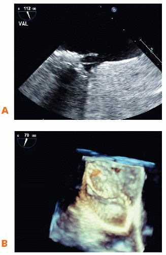
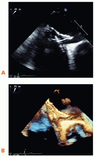
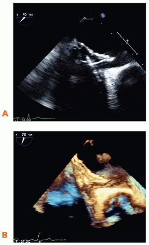
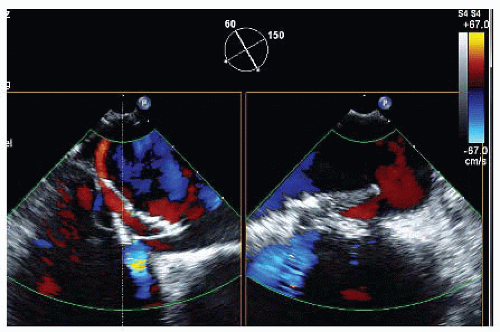
At a 6-month follow-up transesophageal echocardiogram (TEE) after PFO closure, Figures 32-1, 32-2 and 32-3 and Videos 32-1 to 32-3 were obtained to confirm device position.
 Figure 32-3. A. 2D TEE: Long-axis (LAX) view (112°) contrast study: Administration of agitated saline via a cubital vein. B. 3D TEE enface view from the left atrial (LA) side. |
A. The septum secundum is sealed adequately by the left and right atrial discs of the Helex septal occluder
B. No complications after device implantation can be detected
C. A complication after device closure can be verified
D. A residual shunt can be detected
View Answer
ANSWER 1: C. The images (Fig. 32-1 and Video 32-1) demonstrate that the septum secundum is not adequately sealed by the left and right atrial discs; a gap is seen along the septum secundum. The color flow (Fig. 32-2 and Video 32-2) clearly documents a residual shunt in this region, which has to be considered a complication.
 Figure 32-1. A. 2D TEE 45°. B. 3D TEE full-volume acquisition.
Stay updated, free articles. Join our Telegram channel
Full access? Get Clinical Tree
 Get Clinical Tree app for offline access

|

