Fig. 19.1
An illustration of the location of atrial septal defects (ASD). The left panel is a pathological specimen of a normal heart with the anterior surface of the heart removed. Viewed from the right atrium the atrial septum is enclosed within the dotted line. The right panel is a diagrammatic representation of the pathological specimen showing the relative locations of the four types of ASD. Abbreviations: 1 ostium secundum ASD, 2 ostium primum ASD, 3 sinus venosus ASD, 4 coronary sinus ASD, SVC superior vena cava, IVC inferior vena cave, RAA right atrial appendage, CS coronary sinus, TV anterior tricuspid valve leaflet, RV right ventricle, TA tricuspid valve annulus, AVS atrioventricular septum, valve the valve of the inferior vena cava (Eustachian valve), PT pulmonary trunk (pulmonary artery) (Reproduced or adapted from Driscoll, David (2006) Fundamentals of pediatric cardiology. Lippincott Williams & Wilkins, with permission of the author and publisher)
1.
Ostium secundum
2.
Ostium primum
3.
Sinus venosus
4.
Unroofed coronary sinus
The most common form of ASD is the ostium secundum ASD. This occurs in the region of the fossa ovalis. It results from excessive absorption of the septum primum or insufficient development of the septum secundum or both.
Ostium primum ASD is a type of atrioventricular sepal defect (formerly known as “endocardial cushion defects”). It is located in the inferior aspect of the atrial septum contiguous with the tricuspid and mitral valves and will be discussed further in Chap. 25.
Sinus venosus ASDs occur in the posterosuperior aspect of the atrial septum. Frequently there is associated partial anomalous drainage of the right upper pulmonary vein. This vein can drain into the superior vena cava or the right atrium. Resorption of the wall between the vena cava and pulmonary vein results in this type of atrial septal defect. This also explains the anomalous drainage of the right upper pulmonary vein into the right atrium or superior vena cava (SVC) commonly associated with sinus venous atrial septal defects. In fact, technically the atrial septum is not defective, but rather the roof of the pulmonary vein is defective.
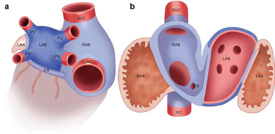
The most uncommon form of ASD is the unroofed coronary sinus. The coronary sinus is apposed to the posterior aspect of the left atrium, but the orifice is in the right atrium. If a hole exists in the roof of the coronary sinus, the coronary sinus and the left atrium will be in continuity, and, therefore the right and left atria will be in communication with each other (Fig. 19.2).