Fig. 27.1
A 50-year-old woman with 15-lb weight loss and upper abdominal pain. (a) Lateral aortogram during inspiration demonstrates a widely patent celiac artery origin (arrow). (b) Lateral aortogram during expiration demonstrates compression of the celiac artery at the level of the median arcuate ligament (arrow)

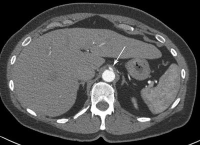
Fig. 27.2
A 38-year-old woman with chronic abdominal pain despite cholecystectomy. Axial images from her CTA identifies a thickened median arcuate ligament (arrow) overlying the celiac artery

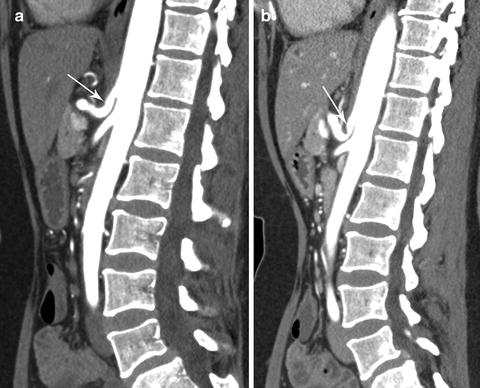
Fig. 27.3
The same 38-year-old woman with sagittal imaging on inspiration and expiration CTA. (a) Widely patent celiac artery (arrow) during inspiration. (b) High-grade stenosis (arrow) of the celiac artery with expiration
In addition, MALS with significant celiac stenosis has been reported as a cause of pancreaticoduodenal artery aneurysm [18–20]. MRA can be used in a similar manner as CTA for patients with a contrast allergy or in children to avoid radiation. However, expiration phase images are impractical with MRA and may need to be combined with ultrasound imaging to identify dynamic changes in the celiac artery [14]. In addition, MRA may not identify intra-abdominal pathology as well as CT scan.
Injection of Celiac Ganglion and Gastric Tonometry
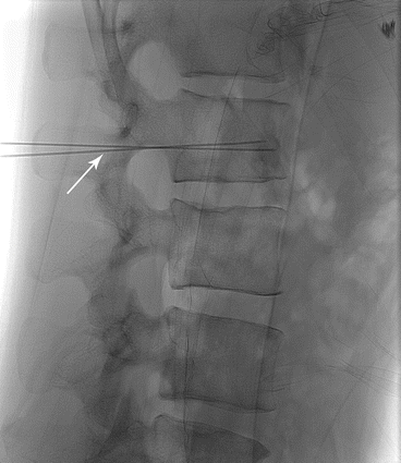
Because median arcuate compression can be found in both symptomatic and asymptomatic patients, imaging alone may not identify patients who will improve after MAL surgical release. The percutaneous celiac ganglion block has been used for many years to control intractable pain for patients with chronic pancreatitis or intra-abdominal cancers. Although permanent block can be obtained by injecting alcohol, a temporary celiac ganglion injection with local anesthetic performed under CT or fluoroscopic guidance (Fig. 27.4) may help to identify patients who would improve from surgical treatment [21]. Although promising, improvement from ganglion block does not absolutely predict future success from MAL surgical release, and further studies are still necessary.


Fig. 27.4
A 25-year-old man with exercise-induced abdominal pain underwent fluoroscopic-guided celiac ganglion temporary block to aid in diagnosis of MALS
Gastric tonometry (See Chap. 6) has also been proposed as a preoperative predictor of success from MAL release. Mensink et al. reported on 43 patients with significant celiac compression of which 30 had ischemia based on gastric exercise tonometry [22]. Of those 30 patients, 29 underwent MAL release and/or revascularization, and 83 % were asymptomatic at 39 months of follow-up. Repeat tonometry in the patients who were asymptomatic was improved in 100 % but only improved in 25 % of patients with persistent symptoms, implicating gastric ischemia an etiology.
Differential Diagnosis
Despite an overlap in symptoms, MALS is distinct from chronic mesenteric ischemia. As opposed to chronic mesenteric ischemia, MALS will not progress to cachexia, bowel gangrene, or death. In addition, MALS typically involves only the celiac artery, although occasionally the ligament can compress the SMA as well. Although initial general symptoms of abdominal pain, postprandial pain, and food fear may lead to the differential diagnosis of both MALS and chronic mesenteric ischemia, careful history and excellent imaging can often easily differentiate between the two diseases.
References
1.
Harjola PT. A rare obstruction of the celiac artery. Ann Chir Gynaecol Fenn. 1963;52:547–50.PubMed
Stay updated, free articles. Join our Telegram channel

Full access? Get Clinical Tree


